Intrinsically photosensitive retinal ganglion cells
Intrinsically photosensitive Retinal Ganglion Cells (ipRGCs), also called photosensitive Retinal Ganglion Cells (pRGC), or melanopsin-containing retinal ganglion cells, are a type of neuron (nerve cell) in the retina of the mammalian eye. While responses to light in mice lacking rods and cone cells were first noted in 1923,[1] they were forgotten, then rediscovered in the early 1990s.[2] The source of these responses was shown to be a special type of retinal ganglion cell, which, unlike other retinal ganglion cells, is intrinsically photosensitive. They are a class of retinal photoreceptors, excited by light even when all influences from the better known photoreceptors (rods and cones) are blocked (either by applying pharmacological agents or by dissociating the ganglion cell from the retina). Photosensitive ganglion cells contain the photopigment melanopsin. The giant retinal ganglion cells of the primate retina are examples of photosensitive ganglion cells.
Overview
Compared to the rods and cones, the ipRGCs respond more sluggishly and signal the presence of light over the long term.[3] They represent a very small subset (~1%) of the retinal ganglion cells (Berson, 2003). Their functional roles are non-image-forming and fundamentally different from those of pattern vision; they provide a stable representation of ambient light intensity. They have at least three primary functions.
- They play a major role in synchronizing circadian rhythms to the 24-hour light/dark cycle, providing primarily length-of-day and length-of night information. They send light information via the retinohypothalamic tract (RHT) directly to the circadian pacemaker of the brain, the suprachiasmatic nucleus of the hypothalamus. The physiological properties of these ganglion cells match known properties of the daily light entrainment (synchronization) mechanism regulating circadian rhythms.
- Photosensitive ganglion cells innervate other brain targets, such as the center of pupillary control, the olivary pretectal nucleus of the midbrain. They contribute to the regulation of pupil size and other behavioral responses to ambient lighting conditions.
- They contribute to photic regulation of, and acute photic suppression of, release of the hormone melatonin from the pineal gland.
Photosensitive ganglion cells are also responsible for the persistence of circadian and pupillary light responses in mammals with degenerated rod and cone photoreceptors, such as humans suffering from retinitis pigmentosa.
Recently photoreceptive ganglion cells have been isolated in humans where, in addition to the above functions shown in other mammals, they have been shown to mediate a degree of light recognition in rodless, coneless subjects suffering with disorders of rod and cone photoreceptors.[4] Work by Farhan H. Zaidi and colleagues showed that photoreceptive ganglion cells may have some visual function in humans.
The photopigment of photoreceptive ganglion cells, melanopsin, is excited by light mainly in the blue portion of the visible spectrum (absorption peaks at ~480 nanometers[5]). The phototransduction mechanism in these cells is not fully understood, but seems likely to resemble that in invertebrate rhabdomeric photoreceptors. Photosensitive ganglion cells respond to light by depolarizing and increasing the rate at which they fire nerve impulses. In addition to responding directly to light, these cells may receive excitatory and inhibitory influences from rods and cones by way of synaptic connections in the retina.
Discovery
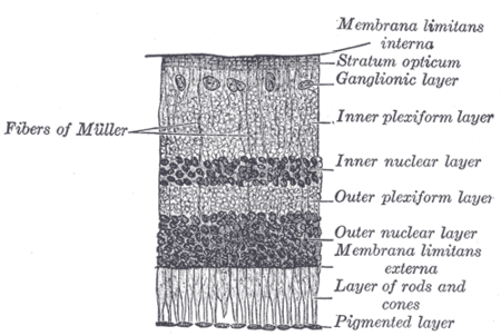
In 1923, Clyde E. Keeler observed that the pupils in the eyes of blind mice he had accidentally bred still responded to light.[6] There was no follow-up at the time. The effect was rediscovered in 1991, when Russell G. Foster and colleagues, including Ignacio Provencio, showed that rods and cones were not necessary for visual drive of the circadian rhythm, i.e. the body's 24-hour biological clock, implying that a third photoreceptor existed in the eye.[7] Later work by Provencio and colleagues showed that this photoresponse was mediated by the photopigment melanopsin, present in the ganglion cell layer of the retina.[8]
The photoreceptors were identified in 2002 by Samer Hattar, David Berson and colleagues, where they were shown to be melanopsin expressing ganglion cells that possessed an intrinsic light response and projected to a number of brain areas involved in non-image forming vision.[9][10]
In 2005, Panda, Melyan, Qiu, and colleagues demonstrated that the melanopsin photopigment was the phototransduction pigment in ganglion cells.[11][12] Dennis Dacey and colleagues showed in a species of Old World monkey that giant ganglion cells expressing melanopsin projected to the lateral geniculate nucleus (LGN).[13][14] Previously only projections to the midbrain (pre-tectal nucleus) and hypothalamus (supra-chiasmatic nuclei, SCN) had been shown. However a visual role for the receptor was still unsuspected and unproven.
Research in humans
Attempts were made to hunt down the receptor in humans, but humans posed special challenges and demanded a new model. Unlike in other animals, researchers could not ethically induce rod and cone loss either genetically or with chemicals so as to directly study the ganglion cells. For many years, only inferences could be drawn about the receptor in humans, though these were at times pertinent.
In 2007, Zaidi and colleagues published their work on rodless, coneless humans, showing that these people retain normal responses to nonvisual effects of light.[4][15] The identity of the non-rod, non-cone photoreceptor in humans was found to be a ganglion cell in the inner retina as shown previously in rodless, coneless models in some other mammals. The work was done using patients with rare diseases that wiped out classic rod and cone photoreceptor function but preserved ganglion cell function.[4][15] Despite having no rods or cones, the patients continued to exhibit circadian photoentrainment, circadian behavioural patterns, melatonin suppression, and pupil reactions, with peak spectral sensitivities to environmental and experimental light that match the melanopsin photopigment. Their brains could also associate vision with light of this frequency. Clinicians and scientists are now seeking to understand the new receptor's role in human diseases and, as discussed below, blindness.[16]
Possible role in conscious sight
Experiments with rodless, coneless humans allowed another possible role for the receptor to be studied. In 2007, a new role was found for the photoreceptive ganglion cell. Zaidi and colleagues showed that in humans the retinal ganglion cell photoreceptor contributes to conscious sight as well as to non-image-forming functions like circadian rhythms, behaviour and pupillary reactions.[4] Since these cells respond mostly to blue light, it has been suggested that they have a role in mesopic vision and that the old theory of a purely duplex retina with rod (dark) and cone (light) light vision was simplistic. Zaidi and colleagues' work with rodless, coneless human subjects hence has also opened the door into image-forming (visual) roles for the ganglion cell photoreceptor.
The discovery that there are parallel pathways for vision was made: one classic rod- and cone-based arising from the outer retina, the other a rudimentary visual brightness detector arising from the inner retina. The latter seems to be activated by light before the other.[4] Classic photoreceptors also feed into the novel photoreceptor system, and colour constancy may be an important role as suggested by Foster.
It has been suggested by the authors of the rodless, coneless human model that the receptor could be instrumental in understanding many diseases including major causes of blindness worldwide such as glaucoma, a disease which affects ganglion cells.
Violet-to-blue light
Most work suggests that the peak spectral sensitivity of the receptor is between 460 and 484 nm. Lockley et al. in 2003[17] showed that 460 nm (violet) wavelengths of light suppress melatonin twice as much as 555 nm (green) light, the peak sensitivity of the photopic visual system. In work by Zaidi, Lockley and co-authors using a rodless, coneless human, it was found that what consciously led to light perception was a very intense 481 nm stimulus; this means that the receptor in visual terms enables some rudimentary vision maximally for blue light.[4]
See also
References
- ↑ Zimmer, Carl (February 2012). "Our Strange, Important, Subconscious Light Detectors". Discover Magazine. Retrieved 2012-02-18.
- ↑ "Seven new Royal Society Fellows". The Medical Sciences Division, University of Oxford. 2008. Retrieved 2010-01-24.
- ↑ Wong KY, Dunn FA, Berson DM (December 2005). "Photoreceptor adaptation in intrinsically photosensitive retinal ganglion cells". Neuron. 48 (6): 1001–10. doi:10.1016/j.neuron.2005.11.016. PMID 16364903.
- 1 2 3 4 5 6 Zaidi FH, Hull JT, Peirson SN, et al. (December 2007). "Short-wavelength light sensitivity of circadian, pupillary, and visual awareness in humans lacking an outer retina". Current Biology. 17 (24): 2122–8. doi:10.1016/j.cub.2007.11.034. PMC 2151130
 . PMID 18082405. Lay summary – Cell Press (December 13, 2007).
 - ↑ Berson DM (August 2007). "Phototransduction in ganglion-cell photoreceptors". Pflügers Archiv. 454 (5): 849–55. doi:10.1007/s00424-007-0242-2. PMID 17351786.
- ↑ Keeler CE (October 1928). "Blind Mice". Journal of Experimental Zoology. 51 (4): 495–508. doi:10.1002/jez.1400510404.
- ↑ Foster RG, Provencio I, Hudson D, Fiske S, De Grip W, Menaker M (July 1991). "Circadian photoreception in the retinally degenerate mouse (rd/rd)". Journal of Comparative Physiology A. 169 (1): 39–50. doi:10.1007/BF00198171. PMID 1941717.
- ↑ Provencio I, Rodriguez IR, Jiang G, Hayes WP, Moreira EF, Rollag MD (January 2000). "A novel human opsin in the inner retina". The Journal of Neuroscience. 20 (2): 600–5. PMID 10632589.
- ↑ Berson DM, Dunn FA, Takao M (February 2002). "Phototransduction by retinal ganglion cells that set the circadian clock". Science. 295 (5557): 1070–3. Bibcode:2002Sci...295.1070B. doi:10.1126/science.1067262. PMID 11834835.
- ↑ Hattar S, Liao HW, Takao M, Berson DM, Yau KW (February 2002). "Melanopsin-containing retinal ganglion cells: architecture, projections, and intrinsic photosensitivity". Science. 295 (5557): 1065–70. Bibcode:2002Sci...295.1065H. doi:10.1126/science.1069609. PMC 2885915. PMID 11834834.
- ↑ Panda S, Nayak SK, Campo B, Walker JR, Hogenesch JB, Jegla T (January 2005). "Illumination of the melanopsin signaling pathway". Science. 307 (5709): 600–4. doi:10.1126/science.1105121. PMID 15681390.
- ↑ Qiu X, Kumbalasiri T, Carlson SM, et al. (February 2005). "Induction of photosensitivity by heterologous expression of melanopsin". Nature. 433 (7027): 745–9. doi:10.1038/nature03345. PMID 15674243.
- ↑ Dacey DM, Liao HW, Peterson BB, et al. (February 2005). "Melanopsin-expressing ganglion cells in primate retina signal colour and irradiance and project to the LGN". Nature. 433 (7027): 749–54. doi:10.1038/nature03387. PMID 15716953.
- ↑ Berson DM (June 2003). "Strange vision: ganglion cells as circadian photoreceptors". Trends in Neurosciences. 26 (6): 314–20. doi:10.1016/S0166-2236(03)00130-9. PMID 12798601.
- 1 2 Coghlan, Andy (2007). "How blind people see sunrise and sunset". New Scientist. 196 (2635–2636): 9. doi:10.1016/S0262-4079(07)63172-8.
- ↑ Schor, Jacob (2008-04-19). "Blue Light and Melatonin" (web page). Morning Light. Retrieved 2008-05-30.
- ↑ Lockley SW, Brainard GC, Czeisler CA (September 2003). "High sensitivity of the human circadian melatonin rhythm to resetting by short wavelength light". The Journal of Clinical Endocrinology and Metabolism. 88 (9): 4502–5. doi:10.1210/jc.2003-030570. PMID 12970330.
| Fibrous tunic (outer) |
| 
 | |||||||||
|---|---|---|---|---|---|---|---|---|---|---|---|
| Uvea/vascular tunic (middle) |
| ||||||||||
| Retina (inner) |
| ||||||||||
| Anatomical regions of the eye |
| ||||||||||
| Other | |||||||||||
External links
- Melanopsin-expressing, Intrinsically Photosensitive Retinal Ganglion Cells, Webvision, University of Utah, USA
- ipRGCs Brown University, Rhode Island, USA