Nervous tissue
| Nervous tissue | |
|---|---|
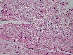 Example of nervous tissue | |
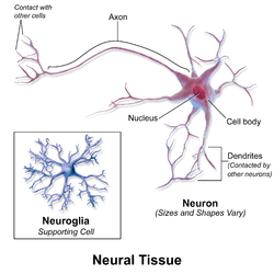 Cells of nervous tissue | |
Nervous tissue or Nerve tissue is the main tissue component of the two parts of the nervous system; the brain and spinal cord of the central nervous system (CNS), and the branching peripheral nerves of the peripheral nervous system (PNS), which regulates and controls bodily functions and activity. It is composed of neurons, or nerve cells, which receive and transmit impulses, and neuroglia, also known as glial cells or more commonly as just glia (from the Greek, meaning glue), which assist the propagation of the nerve impulse as well as providing nutrients to the neuron.
Nervous tissue is made up of different types of nerve cells, all of which have an axon, the long stem-like part of the cell that sends action potential signals to the next cell. Bundles of axons make up the nerves.
Functions of the nervous system are sensory input, integration, control of muscles and glands, homeostasis, and mental activity.
Structure
Nervous tissue is composed of neurons, also called nerve cells, and neuroglial cells. Typically, nervous tissue is categorized into four types of tissue. In the central nervous system (CNS), the tissue types found are grey matter and white matter. In the peripheral nervous system (PNS), the tissue types are nerves and ganglia. The tissue is categorized by its neuronal and neuroglial components.[1]
Components
Neurons are cells with specialized features that allow them to receive and facilitate nerve impulses, or action potentials, across their membrane to the next neuron.[2] They possess a large cell body (soma,perikaryon,cyton), with cell projections called dendrites and an axon. Dendrites are thin, branching projections that receive electrochemical signaling (neurotransmitters) to create a change in voltage in the cell. Axons are long projections that carry the action potential away from the cell body toward the next neuron. The bulb-like end of the axon, called the axon terminal, is separated from the dendrite of the following neuron by a small gap called a synaptic cleft. When the action potential travels to the axon terminal, neurotransmitters are released across the synapse and bind to the post-synaptic receptors, continuing the nerve impulse.[3]
Neurons are classified both functionally and structurally.
Functional classification:[4]
- Sensory neurons (afferent): Relay sensory information in the form of an action potential (nerve impulse) from the PNS to the CNS
- Motor neurons (efferent): Relay an action potential out of the CNS to the proper effector (muscles, glands)
- Interneurons: Cells that form connections between neurons and whose processes are limited to a single local area in the brain or spinal cord
Structural classification:[4]
- Multipolar neurons: Have 3 or more processes coming off the soma (cell body). They are the major neuron type in the CNS and include interneurons and motor neurons.
- Bipolar neurons: Sensory neurons that have two processes coming off the soma, one dendrite and one axon
- Pseudounipolar neurons: Sensory neurons that have one process that splits into two branches, forming the axon and dendrite
- Unipolar brush cells: Are excitatory glutamatergic interneurons that have a single short dendrite terminating in a brush-like tuft of dendrioles. These are found in the granular layer of the cerebellum.
Neuroglia encompasses the non-neural cells in nervous tissue that provide various crucial supportive functions for neurons. They are smaller than neurons, and vary in structure according to their function.[3]
Neuroglial cells are classified as follows:[5]
- Microglial cells: Microglia are macrophage cells that make up the primary immune system for the CNS.[6] They are the smallest neuroglial cell.
- Astrocytes: Star-shaped macroglial cells with many processes found in the CNS. They are the most abundant cell type in the brain, and are intrinsic to a healthy CNS.[7]
- Oligodendrocytes: CNS cells with very few processes. They form myelin sheaths on the axons of a neuron, which are lipid-based insulation that increases the speed at which the action potential, can travel down the axon.[4]
- NG2 glia: CNS cells that are distinct from astrocytes, oligodendrocytes, and microglia, and serve as the developmental precursors of oligodendrocytes[5]
- Schwann cells: The PNS equivalent of oligodendrocytes, they help maintain axons and form myelin sheaths in the PNS.[4]
- Satellite glial cell: Line the surface of neuron cell bodies in ganglia (groups of nerve body cells bundled or connected together in the PNS)[8]
- Enteric glia: Found in the enteric nervous system, within the gastrointestinal tract.[9]
Classification of Tissue
In the Central Nervous System:[10]
- Grey matter is composed of cell bodies, dendrites, unmyelinated axons, protoplasmic astrocytes (astrocyte subtype), satellite oligodendrocytes (non-myelinating oligodendrocyte subtype), microglia, and very few myelinated axons.
- White matter is composed of myelinated axons, fibrous astrocytes, myelinating oligodendrocytes, and microglia.
In the Peripheral Nervous System:[11]
- Ganglion tissue is composed of cell bodies, dendrites, and satellite glial cells.
- Nerves are composed of myelinated and unmyelinated axons, Schwann cells surrounded by connective tissue.
The three layers of connective tissue surrounding each nerve are:[10]
- Endoneurium. Each nerve axon, or fiber is surrounded by the endoneurium, which is also called the endoneurial tube, channel or sheath. This is a thin, delicate, protective layer of connective tissue.
- Perineurium. Each nerve fascicle containing one or more axons, is enclosed by the perineurium, a connective tissue having a lamellar arrangement in seven or eight concentric layers. This plays a very important role in the protection and support of the nerve fibers and also serves to prevent the passage of large molecules from the epineurium into a fascicle.
- Epineurium. The epineurium is the outermost layer of dense connective tissue enclosing the (peripheral) nerve.
Function
The function of nervous tissue is to form the communication network of the nervous system by conducting electric signals across tissue.[12] In the CNS, grey matter, which contains the synapses, is important for information processing. White matter, containing myelinated axons, connects and facilitates nerve impulse between grey matter areas in the CNS.[13] In the PNS, the ganglion tissue, containing the cell bodies and dendrites, contain relay points for nerve tissue impulses. The nerve tissue, containing myelinated axons bundles, carry action potential/nerve impulses.[10]
Clinical significance
Tumours
Neoplasms (tumours) in nervous tissue include:
- Gliomas (glial cell tumors)
- Gliomatosis cerebri, Oligoastrocytoma, Choroid plexus papilloma, Ependymoma, Astrocytoma (Pilocytic astrocytoma, Glioblastoma multiforme), Dysembryoplastic neuroepithelial tumour, Oligodendroglioma, Medulloblastoma, Primitive neuroectodermal tumor
- Neuroepitheliomatous tumors
- Ganglioneuroma, Neuroblastoma, Atypical teratoid rhabdoid tumor, Retinoblastoma, Esthesioneuroblastoma
- Neurofibroma (Neurofibrosarcoma, Neurofibromatosis), Schwannoma, Neurinoma, Acoustic neuroma, Neuroma
References
- ↑ "Peripheral Nervous System". Histology and Virtual Microscopy Learning Resource. University of Michigan Medical School. Retrieved 29 January 2015.
- ↑ Byrne, John; Roberts, James (2004). From Molecules to Networks. California: Academic Press. p. 1. Check date values in:
|access-date=(help); - 1 2 Swenson, Rand. "Review of Clinical and Functional Neuroscience". Dartmouth Medical School. Retrieved 30 January 2015.
- 1 2 3 4 Waymire, Jack. "Organization of Cell Types". Neuroscience Online. The University of Texas Medical School. Retrieved 27 January 2015.
- 1 2 Verkhratsky, Alexi; Butt, Arthur (2013). Glial Physiology and Pathaphysiology (PDF) (First ed.). Chinchester, UK: John Wiley & Sons. p. 76. Retrieved 27 January 2015.
- ↑ Brodal, Per (March 1, 2010). The Central Nervous System: Structure and Function (Fourth ed.). Oxford University Press. p. 19. Retrieved 27 January 2015.
- ↑ Sofroniew, Michael; Vinters, Harry (2009). "Astrocytes: biology and pathology". Acta Neuropathol. 119 (1): abstract. doi:10.1007/s00401-009-0619-8. PMC 2799634
 . PMID 20012068.
 - ↑ M, Hanani (2010). "Satellite glial cells in sympathetic and parasympathetic ganglia: in search of function". Brain Research Review. 64 (2): 1. doi:10.1016/j.brainresrev.2010.04.009. PMID 20441777. Retrieved 27 January 2015.
- ↑ Gershon, Michael; Rothman, Taube (1991). "Enteric Glia". Department of Anatomy and Cell Biology. 4: 195–204. doi:10.1002/glia.440040211.
- 1 2 3 "Neurons and Support Cells". SIU Med. Southern Illinois University School of Medicine. Retrieved 31 January 2015.
- ↑ "Cellular Components of Nervous Tissue" (PDF). RMC faculty. Randolph-Macon College. Retrieved 20 January 2015.
- ↑ "Nervous Tissue". Sidwell School. Retrieved 27 January 2015.
- ↑ Robertson, Sally. "What is Grey Matter". News Medical. AZo Network. Retrieved 30 January 2015.