Negri bodies
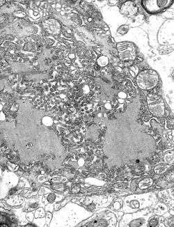
Micrograph with numerous rabies virions (small dark-grey rod-like particles) and Negri bodies, larger pathognomonic cellular inclusions of rabies infection.
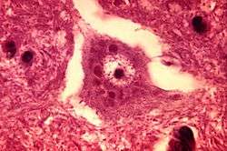
Description: This micrograph depicts the histopathologic changes associated with rabies encephalitis prepared using an H&E stain. Note the Negri bodies, which are cellular inclusions found most frequently in the pyramidal cells of Ammon's horn, and the Purkinje cells of the cerebellum. They are also found in the cells of the medulla and various other ganglia.
Negri bodies are eosinophilic, sharply outlined, pathognomonic inclusion bodies (2–10 µm in diameter) found in the cytoplasm of certain nerve cells containing the virus of rabies, especially in Ammon's horn of the hippocampus. They are also often found in the cerebellar cortex of postmortem brain samples of rabies victims. They consist of ribonuclear proteins produced by the virus.
They are named for Adelchi Negri.[1]
References
External links
This article is issued from Wikipedia - version of the 11/27/2016. The text is available under the Creative Commons Attribution/Share Alike but additional terms may apply for the media files.