Navicular bone
| Navicular bone | |
|---|---|
 The left navicular. Antero-lateral view | |
 The left navicular. Postero-medial view. | |
| Details | |
| Identifiers | |
| Latin | os naviculare |
| TA | A02.5.12.001 |
| FMA | 24499 |
The navicular bone /nəˈvɪkjᵿlər/ is a small bone found in the feet of most mammals.
Human anatomy
The navicular bone in humans is one of the tarsal bones, found in the foot. Its name derives from the human bone's resemblance to a small boat, caused by the strongly concave proximal articular surface. The term navicular bone or hand navicular bone was formerly used for the scaphoid bone,[1] one of the carpal bones of the wrist.
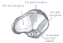
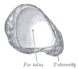
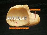
The navicular bone in humans is located on the medial side of the foot, and articulates proximally with the talus, distally with the three cuneiform bones, and laterally with the cuboid.
It is the last of the foot bones to start ossification and does not tend to do so until the end of the third year in girls and the beginning of the fourth year in boys, although a large range of variation has been reported.[2]


The tibialis posterior is the only muscle that attaches to the navicular bone. The main portion of the muscle inserts into the tuberosity of the navicular bone.[3] An accessory navicular bone may be present in 2–14% of the general population.[4][5][6]
Clinical significance
The human navicular is not a commonly broken bone.
Horse anatomy
The horse has a sesamoid bone called the navicular bone, located within the hoof, that lies on the palmar aspect of the coffin joint between the second phalanx and third phalanx (coffin bone). The navicular bone in the horse is supported by the distal sesamoidean impar ligament and two collateral sesamoidean ligaments. The navicular bursa is located between the flexor surface of the navicular bone and the deep digital flexor tendon, which runs between the bursa and the distal phalanx. [7] The central tarsal bone in the hock of the horse is homologous and analogous to the navicular bone of the human foot, and thus the navicular bone in the horse is a different structure from the eponymously labeled bone in humans.[8]
The navicular region is an important structure in relation to lameness, particularly in the front feet, and is involved with a significant disease process called navicular disease or navicular syndrome. Recently much of the original literature concerning navicular disease has been called into question, particularly the significance of radiographic changes as a sole diagnostic criterion.[9] Navicular syndrome may be responsible for as much as 1/3 of all cases of lameness in horses, but radiographic changes in the navicular bone do not always provide a definitive diagnosis. Newer imaging techniques have shown that damage to the soft tissues in the region may be significant contributors to lameness and that multiple causes may result in visible lameness.[7]
See also
| Wikimedia Commons has media related to Navicular bone. |
Notes
- ↑ "Gray's Anatomy, 6b. The Hand. 1. The Carpus. 4". 1918. Retrieved December 2009. Check date values in:
|access-date=(help) - ↑ Louise Scheuer & Sue Black (2004). The Juvenile Skeleton.
- ↑ Bojsen-Møller, Finn; Simonsen, Erik B.; Tranum-Jensen, Jørgen (2001). Bevægeapparatets anatomi [Anatomy of the Locomotive Apparatus] (in Danish) (12th ed.). p. 293. ISBN 978-87-628-0307-7.
- ↑ http://footandankle.mdmercy.com/conditions/flatfeet/navicular.html
- ↑ http://www.wheelessonline.com/ortho/accessory_navicular
- ↑ http://www.macrorad.com/case-reports/symptomatic-accessory-navicular-bone.html
- 1 2 R. Wayne Waguespack, DVM, MS, DACVS R. Reid Hanson, DVM, DACVS, DACVECC (December 2010). "Navicular Syndrome in Equine Patients: Anatomy, Causes, and Diagnosis" (PDF). Surgical Views. Auburn University. Retrieved 11 January 2013.
- ↑ Chapter 32, FRACTURE AND LUXATION OF THE TARSUS AND METATARSUS "Textbook of Small Animal Orthopaedics" written by Charles D. Newton, D.V.M., M.S. and David M. Nunamaker, V.M.D. J.B. Lippincott Company, 1985.
- ↑ Citing Clinical Anatomy and Physiology Laboratory Manual for Veterinary Technicians, Colville, Thomas and Bassert, Joanna M. 2008 Publ. Mosby/Elsevier, Canada. "Navicular Bone - The distal sesamoid bone of the horse. The navicular bone is located deep in the hoof behind the joint between the middle and distal phalanges."
External links
- 3D printable navicular bone model, free download in STL format (Embodi3D.com)