Myelencephalon
| Myelencephalon Afterbrain | |
|---|---|
 | |
| Details | |
| Identifiers | |
| Latin | Myelencephalon |
| MeSH | A08.186.211.132.810.406 |
| NeuroNames | hier-695 |
| TA | A14.1.03.003 |
| FMA | 62004 |
The myelencephalon or afterbrain is the most posterior region of the embryonic hindbrain, from which the medulla oblongata develops.[1]
Development
Neural tube to Myelencephalon
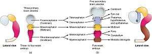
During fetal development, divisions of the neural tube that give rise to the hindbrain (rhombencephalon) and the other primary vesicles (forebrain and midbrain) occur at just 28 days after conception. With the exception of the midbrain, these primary vesicles undergo further differentiation at 5 weeks after conception to form the myelencephalon and the other secondary vesicles.[2]
Myelencephalon to medulla
Final shape differentiation of the myelencephalon into the medulla oblongata can be observed at 20 weeks gestation.[2]
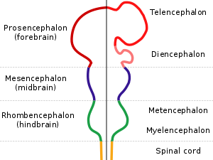
| Neural Tube | Primary Vesicles | Secondary Vesicles | Adult Structures |
| Brain | Forebrain | Telencephalon | Rhinencephalon, Amygdala, Hippocampus, Cerebrum(Cortex), Basal Ganglia,Lateral ventricles |
| Diencephalon | Epithalamus, Thalamus, Hypothalamus, Subthalamus, Pituitary, Pineal, Third ventricle | ||
| Midbrain | Mesencephalon | Tectum, Cerebral peduncle, Pretectum, Cerebral aqueduct | |
| Hindbrain | Metencephalon | Pons, Cerebellum | |
| Myelencephalon | Medulla Oblongata | ||
| Spinal cord | |||
Medulla oblongata
The medulla oblongata is part of the brain stem that serves as the connection of the spinal cord to the brain. It is situated between the pons and the spinal cord.

Function
The medulla oblongata is responsible for several functions of the autonomic nervous system. These functions include:[5]
1) Respiration: monitors the acidity of the blood and sends electrical signals to intercostal muscle tissue to increase their contraction rate in order to oxygenate the blood as needed.
2) Cardiac & Vasomotor Center:[6] monitors and regulates cardiovascular activities by:
- Sympathetic excitation in order to increase cardiac output
- Parasympathetic inhibition of cardiac output
- Affecting blood pressure via vasodilation and vasoconstriction
3) Reflexes
- Coughing
- Sneezing
- Swallowing (palatal)
- Vomiting
- Gagging (pharyngeal)
- Jaw jerk (masseter)
Damage/trauma
Because of its location in the brainstem and its many important roles in the autonomic nervous system, damage to the medulla oblongata is usually fatal.
References
- ↑ "Myelencephalon". Segen's Medical Dictionary. 2011. Retrieved 2015-05-05.
- 1 2 Carlson, Neil R. Foundations of Behavioral Neuroscience.63-65
- ↑ "Neural - Myelencephalon Development - Embryology". embryology.med.unsw.edu.au. Retrieved 2015-05-05.
- ↑ "OpenStax CNX". cnx.org. Retrieved 2015-05-05.
- ↑ Loewy, A. D., & Spyer, K. M. (Eds.). (1990). Central regulation of autonomic functions. Oxford University Press, USA.145-164
- ↑ "Cardiovascular Regulation" (PDF). www.colorado.edu. Retrieved 2015-05-05.