Mossy fiber (hippocampus)
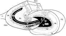
In the hippocampus, granule cells of the dentate gyrus form distinctive unmyelinated axons that project along the mossy fiber pathway to the Cornu Ammonis area 3 (CA3). The axons emerge from the basal portions of the granule cells and pass through the hilus (or polymorphic cell layer) of the dentate gyrus before entering the stratum lucidum of CA3. Granule cell synapses tend to be glutamatergic (e.g. excitatory), though immunohistological data has indicated that some synapses contain neuropeptidergic elements including opiate peptides such as dynorphin and enkephalin. Moreover, there is growing evidence for co-localization of GABAergic and glutamatergic neurotransmitters within mossy fiber terminals, though the functional consequence of this co-localization is unclear.[2]
The pathway was so named by Ramon y Cajal because the axons display varicosities all along their lengths, giving them a "mossy" appearance. They form three morphologically different synaptic terminals, which include the large "mossy terminals", "filopodial extensions" of the mossy terminals, and smaller "en passant" synaptic varicosities.[3] Mossy fibers form multiple synapses with the elaborate dendritic spines of CA3 pyramidal cells in stratum lucidum. These complex spines are known as "thorny excrescences".[4] It has also been shown that the axons of granule cells synapse with dentate gyrus hilar mossy cells and GABAergic interneurons before reaching pyramidal cells in the CA3 region. Thorny excrescences also cover the proximal dendrites of mossy cells. Hilar thorny excrescences are more dense and complex than CA3 ones, and provide the most distinctive feature of mossy cells.[5]
A single mossy fiber projection may make as many as 37 contacts with a single pyramidal cell, but innervates only about a dozen different pyramidal cells. In contrast, a single CA3 pyramidal cell receives input from about 50 different granule cells. It has been shown in rodents that the size of the mossy fiber projections can show large interindividual variations, which are to a large part heritable.[6] In addition, these variations show strong correlations with different types of behavior, mainly, but not exclusively, spatial learning.[7]
The synapses of the mossy fibers contain zinc, which can be stained specifically with a Timm staining.[8]
See also
References
- ↑ Sluyter, Frans; Jamot, Laure; Bertholet, Jean-Yves; Crusio, Wim (2005-04-22). "Prenatal exposure to alcohol does not affect radial maze learning and hippocampal mossy fiber sizes in three inbred strains of mouse". Behavioral and Brain Functions. 1 (1): 5. doi:10.1186/1744-9081-1-5. ISSN 1744-9081.
- ↑ Sandler, R; Smith, AD (1991). "Coexistence of GABA and glutamate in mossy fiber terminals of the primate hippocampus: An ultrastructural study". The Journal of Comparative Neurology. 303 (2): 177–192. doi:10.1002/cne.903030202. PMID 1672874.
- ↑ Acsády, László; Kamondi, A; Sík, A; Freund, T; Buzsáki, G (1998). "GABAergic cells are the major postsynaptic targets of mossy fibers in the rat hippocampus". Journal of Neuroscience. 18 (9): 3386–3403. PMID 9547246.
- ↑ Gonzales, Roberto (2001). "Distribution of thorny excrescences on CA3 pyramidal neurons in the rat hippocampus". Journal of Comparative Neurology. 430 (3): 357–368. doi:10.1002/1096-9861(20010212)430:3<357::aid-cne1036>3.0.co;2-k. PMID 11169473.
- ↑ Amaral, DG; Scharfman, HE; Lavenex, P (2007). "The dentate gyrus: fundamental neuroanatomical organization (dentate gyrus for dummies).". Progress in brain research. 163: 3–22. doi:10.1016/S0079-6123(07)63001-5. PMC 2492885
 . PMID 17765709.
 - ↑ Crusio, WE; Genthner-Grimm, G.; Schwegler, H. (2007). "A quantitative-genetic analysis of hippocampal variation in the mouse". J. Neurogenet. 21 (4): 197–208. doi:10.1080/01677060701715827. PMID 18161583. Retrieved 2009-01-17.
- ↑ Crusio, WE; Schwegler, H. (April 2005). "Learning spatial orientation tasks in the radial-maze and structural variation in the hippocampus in inbred mice". Behav Brain Funct. 1 (1): 3. doi:10.1186/1744-9081-1-3. PMC 1143776. PMID 15916698. Retrieved 2009-01-17.
- ↑ Danscher, G; Zimmer, J (1978). "An improved Timm sulphide silver method for light and electron microscopic localization of heavy metals in biological tissues". Histochemistry. 55 (1): 27–40. doi:10.1007/bf00496691. PMID 76622.