Middle cardiac nerve
| Middle cardiac nerve | |
|---|---|
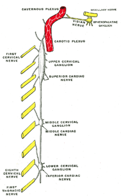 Diagram of the cervical sympathetic. | |
| Details | |
| From | middle cervical ganglion |
| Latin | nervus cardiacus cervicalis medius |
The middle cardiac nerve (great cardiac nerve), the largest of the three cardiac nerves, arises from the middle cervical ganglion, or from the trunk between the middle and inferior ganglia.[1]
On the right side it descends behind the common carotid artery, and at the root of the neck runs either in front of or behind the subclavian artery; it then descends on the trachea, receives a few filaments from the recurrent nerve, and joins the right half of the deep part of the cardiac plexus.
In the neck, it communicates with the superior cardiac and recurrent nerves.
On the left side, the middle cardiac nerve enters the chest between the left carotid and subclavian arteries, and joins the left half of the deep part of the cardiac plexus.
See also
References
- ↑ Power, John Hatch (1860). Anatomy of the arteries of the human body. Fannin and Co. p. 32.
This article is issued from Wikipedia - version of the 10/23/2016. The text is available under the Creative Commons Attribution/Share Alike but additional terms may apply for the media files.