Microspore
Microspores are land plant spores that develop into male gametophytes, whereas megaspores develop into female gametophytes. The male gametophyte gives rise to sperm cells, which are used for fertilization of an egg cell to form a zygote. Microspores are structures that are part of the alternation of generations in many seedless vascular cryptogams, all gymnosperms and all angiosperms. Plants with heterosporous life cycles using microspores and megaspores arose independently in several plant groups during the Devonian period. [1] Microspores are haploid, and are produced from diploid microsporocytes by meiosis. [2]
Seedless vascular plants
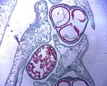
Microsporophylls bear microsporangia containing many microsporocytes that undergo meiosis, producing tiny microspores. Each microspore may become a male gametophyte consisting of a somewhat spherical antheridium within the microspore wall. Either 128 or 256 sperm cells with flagella are produced in each antheridium.[3] In the ferns, the only heterosporous plants are aquatic or semi-aquatic, including the genera Marsilea,Regnellidium, Pilularia, Salvinia, and Azolla. This condition is also known in the lycopod genus Selaginella and in the quillwort genus Isoëtes.
Types of seedless vascular plants:
Gymnosperms
In seed plants the microspores give rise to the pollen grains, and the megaspores are formed within the developing seed. Pollen cones usually develop toward the tips of the lower branches in clusters up to 50 or more. Microsporangia develop in pairs toward the bases of the scales. Each of the microsporocytes in the microsporangia undergoes meiosis, producing four haploid microspores. These develop into pollen grains; each grain consists of four cells and a pair of external air sacs. The air sacs give the pollen grains added buoyancy that could help with wind dispersal. [4]
Types of Gymnosperms:
Angiosperms
As the anther of a flowering plant develops, four patches of tissue differentiate from the main mass of cells. These patches of tissue contain many diploid microsporocyte cells, each of which undergoes meiosis producing a quartet of microspores. Four chambers (pollen sacs) lined with nutritive tapetal cells are visible by the time the microspores are produced. After meiosis, the haploid microspores undergo several changes:
- The microspore divides by mitosis producing two cells. The first of the cells (the generative cell) is small and is formed inside the second larger cell (the tube cell).
- The members of each part of the microspores separate from each other.
- A double-layered wall then develops around each microspore.
These steps occur at relatively the same time and when complete, the microspores have become pollen grains. [5]
See also
References
- ↑ Bateman, R.M.; Dimichele, W.A. (1994). "Heterospory - the most iterative key innovation in the evolutionary history of the plant kingdom.". Biological Reviews of the Cambridge Philosophical Society. 69: 315–417.
- ↑ Bidlack, James E.; Jansky, Shelley H. (2011). Stern's Introductory Plant Biology. New York, NY: McGraw-Hill. ISBN 0-07-304052-5.
- ↑ Bidlack, James E.; Jansky, Shelley H. (2011). Stern's Introductory Plant Biology. New York, NY: McGraw-Hill. ISBN 0-07-304052-5.
- ↑ Bidlack, James E.; Jansky, Shelley H. (2011). Stern's Introductory Plant Biology. New York, NY: McGraw-Hill. ISBN 0-07-304052-5.
- ↑ Bidlack, James E.; Jansky, Shelley H. (2011). Stern's Introductory Plant Biology. New York, NY: McGraw-Hill. ISBN 0-07-304052-5.
| Subdisciplines | |||||||||||||||||||
|---|---|---|---|---|---|---|---|---|---|---|---|---|---|---|---|---|---|---|---|
| Plant groups | |||||||||||||||||||
| |||||||||||||||||||
| |||||||||||||||||||
| Plant growth and habit | |||||||||||||||||||
| Reproduction | |||||||||||||||||||
| Plant taxonomy | |||||||||||||||||||
| Practice | |||||||||||||||||||
| |||||||||||||||||||
| |||||||||||||||||||