Mesonephric duct
| Mesonephric duct | |
|---|---|
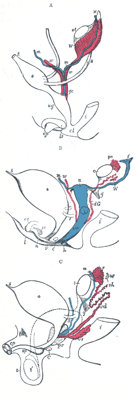
 Urogenital sinus of female human embryo of eight and a half to nine weeks old. | |
 Transverse section of a chick embryo of forty-five hours' incubation. | |
| Details | |
| Carnegie stage | 11 |
| Days | 28 |
| Precursor | intermediate mesoderm |
| Gives rise to | vas deferens, seminal vesicles, epididymis |
| Identifiers | |
| Latin | ductus mesonephricus; ductus Wolffi |
| Code | TE E5.6.2.0.0.0.4 |
The mesonephric duct (also known as Wolffian duct, archinephric duct, Leydig's duct and nephric duct) is a paired organ found in mammals including humans during embryogenesis. Wolffian structures are male urogenital structures that include the epididymis, vas deferens, and seminal vesicles that differentiate from this structure.
The mesonephric duct connects the primitive kidney, the mesonephros, to the cloaca and serves as the anlage for certain male reproductive organs.
Structure
The mesonephric duct connects the primitive kidney, the mesonephros, to the cloaca and serves as the anlage for certain male reproductive organs.
Development
In both the male and the female the mesonephric duct develops into the trigone of urinary bladder, a part of the bladder wall. However, further development differentiates between the sexes in the development of the urinary and reproductive organs.
Male
In a male, it develops into a system of connected organs between the efferent ducts of the testis and the prostate, namely the epididymis, the vas deferens, and the seminal vesicle. The prostate forms from the urogenital sinus and the efferent ducts form from the mesonephric tubules.
For this it is critical that the ducts are exposed to testosterone during embryogenesis. Testosterone binds to and activates androgen receptor, affecting intracellular signals and modifying the expression of numerous genes.[1]
In the mature male, the function of this system is to store and mature sperm, and provide accessory semen fluid.
Female
In the female, with the absence of anti-Müllerian hormone secretion by the Sertoli cells and subsequent Müllerian apoptosis, the Wolffian duct regresses, and inclusions may persist. The epoophoron and Skene's glands may be present. Also, lateral to the wall of the vagina a Gartner's duct or cyst could develop as a remnant.
Function
Sexual differentiation
History
It is named after Caspar Friedrich Wolff who described the mesonephros and its ducts in his dissertation in 1759.[2]
Additional images
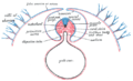 Diagram of a transverse section, showing the mode of formation of the amnion in the chick.
 Reconstruction of a human embryo of 17 mm.
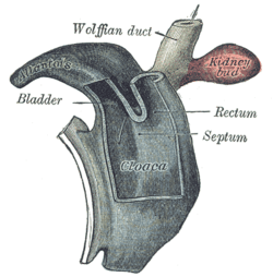 Cloaca of human embryo from twenty-five to twenty-seven days old.
 Broad ligament of adult, showing epoophoron.
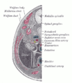 Transverse section of human embryo eight and a half to nine weeks old.
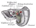 Tail end of human embryo twenty-five to twenty-nine days old.
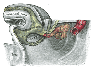 Tail end of human embryo thirty-two to thirty-three days old.
 Tail end of human embryo; from eight and a half to nine weeks old.
 Primitive kidney and bladder, from a reconstruction.
See also
- Fetal genital development
- List of homologues of the human reproductive system
- Masculinization
- Müllerian duct
- Sexual differentiation
References
- ↑ Hannema SE, Print CG, Charnock-Jones DS, Coleman N, Hughes IA (2006). "Changes in gene expression during Wolffian duct development". Horm. Res. 65 (4): 200–9. doi:10.1159/000092408. PMID 16567946.
- ↑ synd/2845 at Who Named It?
External links
- MedicalMnemonics.com: 1266
- How the Body Works / Sex Development / Sexual Differentiation / Duct Differentiation - The Hospital for Sick Children (GTA - Toronto, Ontario, Canada)