Medial arcuate ligament
| Medial arcuate ligament | |
|---|---|
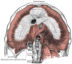 The diaphragm. Under surface. (Med. arcuate ligament visible at bottom center left.) | |
| Details | |
| Identifiers | |
| Latin | ligamentum arcuatum mediale |
| TA | A04.4.02.006 |
| FMA | 58282 |
The medial arcuate ligament (also medial lumbocostal arch and internal arcuate ligament) is a tendinous fascia that arches over the psoas major muscle as it passes through the diaphragm.
Structure
The medial arcuate ligament is an arch in the fascia covering the upper part of the psoas major. It is attached to the side of the body of the first or second lumbar vertebra; near cervical and thoracal vertebra, laterally, it is fixed to the front of the transverse process of the first and, sometimes also, to that of the second lumbar vertebra.
It lies between the lateral arcuate ligament and the midline median arcuate ligament.
The sympathetic chain enters the abdomen by passing deep into this ligament.
See also
References
This article incorporates text in the public domain from the 20th edition of Gray's Anatomy (1918)
External links
- Anatomy figure: 40:04-04 at Human Anatomy Online, SUNY Downstate Medical Center
- posteriorabdomen at The Anatomy Lesson by Wesley Norman (Georgetown University) (posteriorabdmus&nerves)