Maxillary artery
| Maxillary artery | |
|---|---|
 Plan of branches of maxillary artery. ("Internal maxillary" is horizontal at left center.) | |
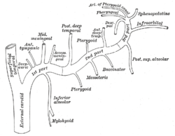 Plan of branches of maxillary artery. | |
| Details | |
| Precursor | aortic arch 1 |
| Source | external carotid artery |
| Branches |
1st part: anterior tympanic - deep auricular - middle meningeal - superior tympanic - accessory meningeal - inferior alveolar 2nd part: Posterior deep temporal artery - Pterygoid branches - masseteric - buccinator - Anterior deep temporal artery 3rd part: posterior superior alveolar - infraorbital - descending palatine - artery of the pterygoid canal - sphenopalatine |
| Vein | maxillary veins |
| Identifiers | |
| Latin | arteria maxillaris |
| TA | A12.2.05.053 |
| FMA | 49675 |
The maxillary artery supplies deep structures of the face. It branches from the external carotid artery just deep to the neck of the mandible.
Structure
The maxillary artery, the larger of the two terminal branches of the external carotid artery, arises behind the neck of the mandible, and is at first imbedded in the substance of the parotid gland; it passes forward between the ramus of the mandible and the sphenomandibular ligament, and then runs, either superficial or deep to the lateral pterygoid muscle, to the pterygopalatine fossa.
It supplies the deep structures of the face, and may be divided into mandibular, pterygoid, and pterygopalatine portions.
First portion
The first or mandibular portion (or bony portion) passes horizontally forward, between the neck of the mandible and the sphenomandibular ligament, where it lies parallel to and a little below the auriculotemporal nerve; it crosses the inferior alveolar nerve, and runs along the lower border of the lateral pterygoid muscle.
Branches include:
- Deep auricular artery
- Anterior tympanic artery
- Middle meningeal artery
- Inferior alveolar artery which gives off its mylohyoid branch just prior to entering the mandibular foramen
- Accessory meningeal artery
Second portion
The second or pterygoid portion (or muscular portion) runs obliquely forward and upward under cover of the ramus of the mandible and insertion of the temporalis, on the superficial (very frequently on the deep) surface of the lateral pterygoid muscle; it then passes between the two heads of origin of this muscle and enters the fossa.
Branches include:
- Masseteric artery
- Pterygoid branches
- Deep temporal arteries (anterior and posterior)
- Buccal artery
Third portion
The third or pterygomaxillary portion lies in the pterygopalatine fossa in relation with the pterygopalatine ganglion. This is considered the terminal branch of the maxillary artery.
Branches include:
- Sphenopalatine artery (Nasopalatine artery is the terminal branch of the Maxillary artery)
- Descending palatine artery (Greater palatine artery and lesser palatine artery)
- Infraorbital artery
- Posterior superior alveolar artery
- Artery of pterygoid canal
- Pharyngeal artery
- Middle superior alveolar artery (a branch of the infraorbital artery)
- Anterior superior alveolar arteries (a branch of the infraorbital artery)
Nomenclature
- Formerly, the term "external maxillary artery" was used to describe what is now known as the facial artery (per Terminologia anatomica.) Currently, the term "external maxillary artery" is less commonly used, and the terms "internal maxillary artery" and "maxillary artery" are equivalent.
Additional images
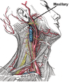 Superficial dissection of the right side of the neck, showing the carotid and subclavian arteries. Origin of maxillary artery is labeled.
 Mandible. Outer surface. Side view.
 Lateral head anatomy detail
 Head anatomy anterior view
 Maxillary artery
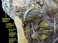 Maxillary artery
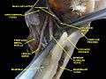 Infratemporal fossa. Lingual and inferior alveolar nerve. Deep dissection. Anterolateral view
References
This article incorporates text in the public domain from the 20th edition of Gray's Anatomy (1918)
External links
- lesson4 at The Anatomy Lesson by Wesley Norman (Georgetown University) (parotid4)
- Anatomy photo:27:12-0101 at the SUNY Downstate Medical Center - "Infratemporal Fossa: Branches of the Maxillary Artery"
- MedicalMnemonics.com: 935
- Overview at tufts.edu