Mandibular notch
| Mandibular notch | |
|---|---|
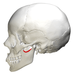 Position of mandibular notch in skull, shown in red. | |
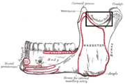
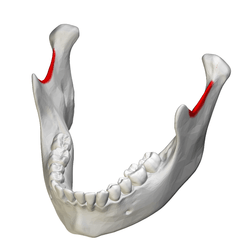 Position of mandibular notch in mandible, shown in red. | |
| Details | |
| Identifiers | |
| Latin | incisura mandibulae |
| TA | A02.1.15.034 |
| FMA | 59481 |
The upper border of the ramus of mandible is thin, and is surmounted by two processes, the coronoid process anteriorly and the condyloid process posteriorly, separated by a deep concavity, the mandibular notch, or sigmoid notch. It allows the passage of the masseteric nerve (a branch of the mandibular nerve (V3) division of the trigeminal nerve), masseteric artery and masseteric vein.
Additional images
|
References
This article incorporates text in the public domain from the 20th edition of Gray's Anatomy (1918)
External links
| Wikimedia Commons has media related to Mandibular notch. |
- Anatomy image: skel/mandible2 at Human Anatomy Lecture (Biology 129), Pennsylvania State University
- Diagram at unc.edu
This article is issued from Wikipedia - version of the 6/9/2015. The text is available under the Creative Commons Attribution/Share Alike but additional terms may apply for the media files.