Magnocellular cell
| Magnocellular cell | |
|---|---|
| Identifiers | |
| NeuroLex ID | Magnocellular cell |
Magnocellular cells, also called M-cells or magnocellular retinal cells, are neurons located within the magnocellular layer of the lateral geniculate nucleus of the thalamus. The cells are part of the visual system. They are termed "magnocellular" since they are characterized by their relatively large size compared to parvocellular cells. M-cells detect the "where" properties of visual information.
Visual system
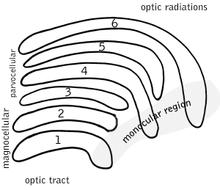
Magnocellular cells are primarily concerned with visual perception.[1] In particular these cells are responsible for resolving motion and coarse outlines. The M-cells receive their information from the axons of parasol cells exiting the optic tract. The M-cells are large, fast-conducting neurons with their cell bodies located in the 2 ventral magnocellular layers of the lateral geniculate nucleus. The information they carry is sent through the optic radiations to the visual cortices, possibly after editing and gating by visual cortex exerting top-down control. The M-cells feed more to the parietal cortices, in the dorsal "where" stream, than the temporal cortices, destination of the ventral "what" stream. This is consistent with their coding of movement and edges as opposed to fine detail. This system of cells operates with great speed at the expense of detail.
See also
References
- ↑ Xu X, Ichida JM, Allison JD, Boyd JD, Bonds AB, Casagrande VA (February 2001). "A comparison of koniocellular, magnocellular and parvocellular receptive field properties in the lateral geniculate nucleus of the owl monkey (Aotus trivirgatus)". J. Physiol. (Lond.). 531 (Pt 1): 203–18. doi:10.1111/j.1469-7793.2001.0203j.x. PMC 2278453
 . PMID 11179404.
External links
| Related | ||
|---|---|---|