Magnetic resonance imaging of the brain
| MRI of brain and brain stem | |
|---|---|
| Diagnostics | |
 Brain MRI | |
| ICD-10-PCS | |
| ICD-9-CM | 88.91 |
| OPS-301 code | 3-800, 3-820 |
Magnetic resonance imaging (MRI) of the nervous system uses magnetic fields and radio waves to produce high quality two- or three-dimensional images of nervous system structures without use of ionizing radiation (X-rays) or radioactive tracers.
History
The first MR images of a human brain were obtained in 1978 by two groups of researchers at EMI Laboratories led by Ian Robert Young and Hugh Clow.[1] In 1986, Charles L. Dumoulin and Howard R. Hart at General Electric developed MR angiography[2] and Denis Le Bihan, obtained the first images and later patented diffusion MRI.[3] In 1990, Seiji Ogawa at AT&T Bell labs recognized that oxygen-depleted blood with dHb was attracted to a magnetic field, and discovered the technique that underlies Functional Magnetic Resonance Imaging (fMRI).[4] In 1997, Jürgen R. Reichenbach, E. Mark Haacke and coworkers at Washington University developed Susceptibility weighted imaging.[5] The first study of the human brain at 3.0 T was published in 1994,[6] and in 1998 at 8 T.[7] Studies of the human brain have been performed at up to 9.4 T.[8] Paul Lauterbur and Sir Peter Mansfield were awarded the 2003 Nobel Prize in Physiology or Medicine for their discoveries concerning MRI.

Applications
One advantage of MRI of the brain over computed tomography of the head is better tissue contrast,[9] and it has fewer artifacts than CT when viewing the brainstem. MRI is also superior for pituitary imaging.[10] It may however be less effective at identifying early cerebritis.[11]
In the case of a concussion, an MRI should be avoided unless there are progressive neurological symptoms, focal neurological findings or concern of skull fracture on exam.[12]
In analysis of the fetal brain, MRI provides more information about gyration than ultrasound.[13]
A number of different imaging modes can be used with imaging the nervous system:
- T1: Cerebrospinal fluid is dark. T1 weighting is useful for visualizing normal anatomy.
- T2: CSF is light, but fat (and thus white matter) is darker than with T1. T2 is useful for visualizing pathology.[14]
- PD (proton density): CSF has a relatively high level of protons, making CSF appear bright. Gray matter is brighter than white matter.[15]
- FLAIR: useful for evaluation of white matter plaques near the ventricles.[16] It is useful in identifying demyelination.[17]
See also
Gallery
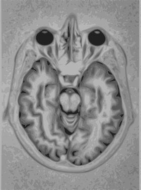
 T1 (note CSF is dark)
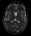 Normal axial T2-weighted MR image of the brain.
| Wikimedia Commons has media related to Magnetic resonance imaging of the brain. |
References
- ↑ "Britain's brains produce first NMR scans". New Scientist: 588. 1978.
- ↑ "Blood-flow checker". Popular Science: 12. 1987.
- ↑ Le Bihan, D; Breton E. (1987). "Method to Measure the Molecular Diffusion and/or Perfusion Parameters of Live Tissue". US Patent # 4,809,701.
- ↑ Faro, Scott H.; Mohamed, Feroze B (2010-01-15). Bold fMRI. a guide to functional imaging for neuroscientists. Springer. ISBN 978-1-4419-1328-9. Retrieved 10 June 2015.
- ↑ Reichenbach JR, Venkatesan R, Schillinger DJ, Kido DK, and Haacke EM (1997). "Small vessels in the human brain: MR venography with deoxyhemoglobin as an intrinsic contrast agent". Radiology. 204 (1): 272–277. doi:10.1148/radiology.204.1.9205259. PMID 9205259.
- ↑ Mansfield P, Coxon R, Glover P (May 1994). "Echo-planar imaging of the brain at 3.0: first normal volunteer results.". J Comput Assist Tomogr. 18 (3): 339–43. PMID 8188896.
- ↑ Robitaille PM, Abduljalil AM, Kangarlu A, Zhang X, Yu Y, Burgess R, Bair S, Noa P, Yang L, Zhu H, Palmer B, Jiang Z, Chakeres DM, Spigos D (Oct 1998). "Human magnetic resonance imaging at 8 T.". NMR Biomed. 11 (6): 263–5. doi:10.1002/(SICI)1099-1492(199810)11:6<263::AID-NBM549>3.0.CO;2-0. PMID 9802467.
- ↑ Vaughan T, DelaBarre L, Snyder C, Tian J, Akgun C, Shrivastava D, Liu W, Olson C, Adriany G, Strupp J, Andersen P, Gopinath A, van de Moortele PF, Garwood M, Ugurbil K (December 2006). "9.4T human MRI: preliminary results". Magn Reson Med. 56 (6): 1274–82. doi:10.1002/mrm.21073. PMID 17075852.
- ↑ Ebel, Klaus-Dietrich; Benz-Bohm, Gabriele (1999). Differential diagnosis in pediatric radiology. Thieme. pp. 538–. ISBN 978-3-13-108131-5. Retrieved 18 July 2011.
- ↑ Bradley, William G.; Brant-Zawadzki, Michael; Cambray-Forker, Jane (2001-01-15). MRI of the brain. Surendra Kumar. ISBN 978-0-7817-2568-2. Retrieved 24 July 2011.
- ↑ Roos, Karen L.; Tunkel, Allan R. (2010). Bacterial infections of the central nervous system. Elsevier Health Sciences. pp. 69–. ISBN 978-0-444-52015-9. Retrieved 18 July 2011.
- ↑ American Medical Society for Sports Medicine (24 April 2014), "Five Things Physicians and Patients Should Question", Choosing Wisely: an initiative of the ABIM Foundation, American Medical Society for Sports Medicine, retrieved 29 July 2014
- ↑ Garel, Cathérine (2004). MRI of the fetal brain: normal development and cerebral pathologies. Springer. ISBN 978-3-540-40747-8. Retrieved 24 July 2011.
- ↑ Butler, Paul; Mitchell, Adam W. M.; Ellis, Harold (2007-11-19). Applied Radiological Anatomy for Medical Students. Cambridge University Press. pp. 12–. ISBN 978-0-521-81939-8. Retrieved 18 July 2011.
- ↑ Tofts, Paul (2005-09-01). Quantitative MRI of the Brain: Measuring Changes Caused by Disease. John Wiley and Sons. pp. 86–. ISBN 978-0-470-86949-9. Retrieved 18 July 2011.
- ↑ Chowdhury, Rajat; Wilson, Iain; Rofe, Christopher; Lloyd-Jones, Graham (2010-04-19). Radiology at a Glance. John Wiley and Sons. pp. 95–. ISBN 978-1-4051-9220-0. Retrieved 18 July 2011.
- ↑ Granacher, Robert P. (2007-12-20). Traumatic brain injury: methods for clinical and forensic neuropsychiatric assessment. CRC Press. pp. 247–. ISBN 978-0-8493-8138-6. Retrieved 18 July 2011.