Lyssavirus
| Lyssavirus | |
|---|---|
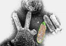 | |
| Colored transmission electron micrograph of Australian bat lyssavirus. The bullet-like objects are the virions, and some of them are budding off from a cell. | |
| Virus classification | |
| Group: | Group V ((-)ssRNA) |
| Order: | Mononegavirales |
| Family: | Rhabdoviridae |
| Genus: | Lyssavirus |
| Type species | |
| Rabies lyssavirus | |
| Species | |
| |
Lyssavirus is a genus of RNA viruses in the family Rhabdoviridae, order Mononegavirales. Humans, mammals, and vertebrates serve as natural hosts.[1][2] The genus Lyssavirus (from Lyssa, the Greek goddess of madness, rage, and frenzy) includes the rabies virus traditionally associated with the disease.
Taxonomy
| Genus | Species | Virus (Abbreviation) |
| Lyssavirus | Aravan lyssavirus | Aravan virus (ARAV) |
| Australian bat lyssavirus | Australian bat lyssavirus (ABLV) | |
| Bokeloh bat lyssavirus | Bokeloh bat lyssavirus (BBLV) | |
| Duvenhage lyssavirus | Duvenhage virus (DUVV) | |
| European bat 1 lyssavirus | European bat lyssavirus 1 (EBLV-1) | |
| European bat 2 lyssavirus | European bat lyssavirus 2 (EBLV-2) | |
| Ikoma lyssavirus | Ikoma lyssavirus (IKOV) | |
| Irkut lyssavirus | Irkut virus (IRKV) | |
| Khujand lyssavirus | Khujand virus (KHUV) | |
| Lagos bat lyssavirus | Lagos bat virus (LBV) | |
| Mokola lyssavirus | Mokola virus (MOKV) | |
| Rabies lyssavirus* | rabies virus (RABV) | |
| Shimoni bat lyssavirus | Shimoni bat virus (SHIBV) | |
| West Caucasian bat lyssavirus | West Caucasian bat virus (WCBV) | |
Table legend: "*" denotes type species.
Virology
Structure
Lyssavirions are enveloped, with bullet shaped geometries. These virions are about 75 nm wide and 180 nm long.[1] Lyssavirions have helical symmetry, so their infectious particles are approximately cylindrical in shape. This is typical of plant-infecting viruses. Virions of human-infecting viruses more commonly have cubic symmetry and take shapes approximating regular polyhedra.
The structure consists of a spiked outer envelope, a middle region consisting of matrix protein M, and an inner ribonucleocapsid complex region, consisting of the genome associated with other proteins.
Genome
Lyssavirus genomes consist of a negative-sense, single-stranded RNA molecule that encodes five viral proteins: polymerase L, matrix protein M, phosphoprotein P, nucleoprotein N, and glycoprotein G. Genomes are linear, around 11kb in length.[1]
Based on recent phylogenetic evidence, lyssaviruses are categorized into seven major species. In addition, five species recently have been discovered: West Caucasian bat virus, Aravan virus, Khujand virus, Irkut virus and Shimoni bat virus.[4][5] The major species include rabies virus (species 1), Lagos bat virus (species 2), Mokola virus (species 3), Duvenhage virus (species 4), European Bat lyssaviruses type 1 and 2 (species 5 and 6), and Australian bat lyssavirus (species 7).[6]
Based on biological properties of the viruses, these species are further subdivided into phylogroups 1 and 2. Phylogroup 1 includes genotypes 1, 4, 5, 6, and 7, while phylogroup 2 includes genotypes 2 and 3. The nucleocapsid region of lyssavirus is fairly highly conserved from genotype to genotype across both phylogroups; however, experimental data have shown the lyssavirus strains used in vaccinations are only from the first species(i.e. classic rabies).[6]
| Genus | Structure | Symmetry | Capsid | Genomic Arrangement | Genomic Segmentation |
|---|---|---|---|---|---|
| Lyssavirus | Bullet-shaped | Enveloped | Linear | Monopartite |
Life Cycle
Viral replication is cytoplasmic. Entry into the host cell is achieved by attachment of the viral G glycoproteins to host receptors, which mediates clathrin-mediated endocytosis. Replication follows the negative stranded RNA virus replication model. Negative stranded RNA virus transcription, using polymerase stuttering is the method of transcription. The virus exits the host cell by budding, and tubule-guided viral movement. Human, mammals, and vertebrates serve as the natural host. Transmission routes are zoonosis and bite.[1]
| Genus | Host Details | Tissue Tropism | Entry Details | Release Details | Replication Site | Assembly Site | Transmission |
|---|---|---|---|---|---|---|---|
| Lyssavirus | Humans; mammals | Neurons | Clathrin-mediated endocytosis | Budding | Cytoplasm | Cytoplasm | Zoonosis; animal bite |
Epidemiology
Classic rabies virus, is prevalent throughout most of the world and can be carried by any warm blooded mammal. The other lyssaviruses have much less diversity in carriers. Only select hosts can carry each of the viral species. Also, these other species are particular only to a specific geographic area. Bats are known to be an animal vector for all identified lyssaviruses except the Mokola virus.[7]
See also
References
- 1 2 3 4 "Viral Zone". ExPASy. Retrieved 15 June 2015.
- ↑ ICTV. "Virus Taxonomy: 2014 Release". Retrieved 15 June 2015.
- ↑ Afonso, Claudio L.; Amarasinghe, Gaya K.; Bányai, Krisztián; Bào, Yīmíng; Basler, Christopher F.; Bavari, Sina; Bejerman, Nicolás; Blasdell, Kim R.; Briand, François-Xavier (2016-08-01). "Taxonomy of the order Mononegavirales: update 2016". Archives of Virology. 161 (8): 2351–2360. doi:10.1007/s00705-016-2880-1. ISSN 1432-8798. PMC 4947412
 . PMID 27216929.
 - ↑ Virus Taxonomy: 2013 Release. ictvonline.org
- ↑ Kuzmin, I.; Hughes, G.; Botvinkin, A.; Orciari, L.; Rupprecht, C. (2005). "Phylogenetic relationships of Irkut and West Caucasian bat viruses within the genus and suggested quantitative criteria based on the N gene sequence for lyssavirus genotype definition". Virus Research. 111 (1): 28–25. doi:10.1016/j.virusres.2005.03.008. PMID 15896400.
- 1 2 Badrane, H.; Bahloul, C.; Perrin, P.; Tordo, N. (2001). "Evidence of Two Lyssavirus Phylogroups with Distinct Pathogenicity and Immunogenicity". Journal of Virology. 75 (7): 3268–3276. doi:10.1128/JVI.75.7.3268-3276.2001. PMC 114120. PMID 11238853.
- ↑ WHO Rabnet/CDC Map Production (2008). "Rabies, countries or areas at risk". World Health Organization.
See also
- Baynard, Ashley C.; Hayman, D.; Johnson, N.; McElhinney, L.; Fooks, A.R. (2011). "Bats and Lyssaviruses". In Jackson, Alan C. Research Advances in Rabies. Advances in Virus Research. 79. Elsevier. ISBN 978-0-12-387040-7.
- Botvinkin; Poleschuk, E. M.; Kuzmin, I. V.; Borisova, T. I.; Gazaryan, S. V.; Yager, P.; Rupprecht, C. E. (2003). "Novel Lyssaviruses Isolated from Bats in Russia". Emerging Infectious Diseases. 9 (12): 1623–1625. doi:10.3201/eid0912.030374. PMC 3034350. PMID 14720408.
- Arai; Kuzmin, I. V.; Kameoka, Y.; Botvinkin, A. D. (2003). "New Lyssavirus Genotype from the Lesser Mouse-eared Bat (Myotis blythi), Kyrghyzstan". Emerging Infectious Diseases. 9 (3): 333–337. doi:10.3201/eid0903.020252. PMC 2958534. PMID 12643828.
- World Health Organization (2005). WHO Expert Consulation on Rabies (PDF). WHO technical report series. Geneva, Switzerland: World Health Organization. ISBN 92-4-120931-3.
External links
| Wikimedia Commons has media related to Lyssavirus. |