Lumbricals of the hand
| Lumbricals of the hand | |
|---|---|
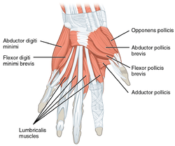
.png) The muscles of the left hand. Palmar surface. (1st lumbricalis labeled at bottom right of muscular group.) | |
| Details | |
| Origin | flexor digitorum profundus |
| Insertion | extensor expansion |
| Artery | superficial palmar arch, common palmar digital arteries, deep palmar arch, dorsal digital artery |
| Nerve | 3rd and 4th deep branch of ulnar nerve, 1st and 2nd median nerve |
| Actions | flex metacarpophalangeal joints, extend interphalangeal joints |
| Identifiers | |
| Latin | musculi lumbricales manus |
| TA | A04.6.02.065 |
| FMA | 37385 |
The lumbricals are intrinsic muscles of the hand that flex the metacarpophalangeal joints and extend the interphalangeal joints.[1]
Structure
There are four of these small, worm-like muscles on each hand. These muscles are unusual in that they do not attach to bone. Instead, they attach proximally to the tendons of flexor digitorum profundus and distally to the extensor expansions.[1]
| # | Form | Origin | Insertion |
| First | unipennate | It originates from the radial side of the most radial tendon of the flexor digitorum profundus (corresponding to the index finger). | It passes posteriorly along the radial side of the index finger to insert on the extensor expansion near the metacarpophalangeal joint. |
| Second | unipennate | It originates from the radial side of the second most radial tendon of the flexor digitorum profundus (which corresponds to the middle finger). | It passes posteriorly along the radial side of the middle finger and inserts on the extensor expansion near the metacarpophalangeal joint. |
| Third | bipennate | One head originates on the radial side of the flexor digitorum profundus tendon corresponding to the ring finger, while the other originates on the ulnar side of the tendon for the middle finger. | The muscle passes posteriorly along the radial side of the ring finger to insert on its extensor expansion. |
| Fourth | bipennate | One head originates on the radial side of the flexor digitorum profundus tendon corresponding to the little finger, while the other originates on the ulnar side of the tendon for the ring finger. | The muscle passes posteriorly along the radial side of the little finger to insert on its extensor expansion. |
Innervation
The first and second lumbricals (the most radial two) are innervated by the median nerve. The third and fourth lumbricals (most ulnar two) are innervated by the deep branch of the ulnar nerve.
This is the usual innervation of the lumbricals (occurring in 60% of individuals). However 1:3 (median:ulnar - 20% of individuals) and 3:1 (median:ulnar - 20% of individuals) also exist. The lumbrical innervation always follows the innervation pattern of the associated muscle unit of flexor digitorum profundus (i.e. if the muscle units supplying the tendon to the middle finger are innervated by the median nerve, the second lumbrical will also be innervated by the median nerve).[2]
Blood supply
There are four separate sources of blood supply for these muscles: the superficial palmar arch, the common palmar digital artery, the deep palmar arch, and the dorsal digital artery.
Actions
The lumbrical muscles, with the help of the interosseous muscles, simultaneously flex the metacarpophalangeal joints while extending both interphalangeal joints of the digit on which it inserts. The lumbricals are used during an upstroke in writing.
Other lumbricals
There are also lumbrical muscles of the foot that have a similar action, though these are of less clinical concern.
Additional images
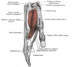 Tendons of forefinger and vincula tendina
 Lumbricals of the hand
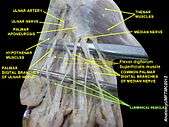 Lumbricals of the hand
 Lumbricals muscle
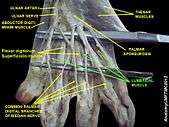 Lumbricals muscle
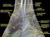 Lumbricals muscle
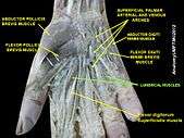 Lumbricals muscle
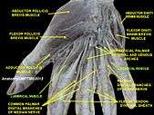 Lumbricals muscle
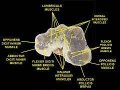 Muscles of hand. Cross section.
 Wrist joint. Deep dissection.Anterior, palmar, view.
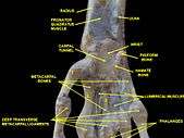 Wrist joint. Deep dissection.Anterior, palmar, view.
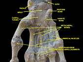 Wrist joint. Deep dissection.Anterior, palmar, view.
Notes
- 1 2 Gosling et al. 2008, p. 97
- ↑ Last's Anatomy - Regional and Applied, 10th ed. Chummy S. Sinnatamby, pg. 64 and pg. 82.
References
- Gosling, J.A.; Harris, P.F.; Humpherson, J.R.; Whitmore, I.; Willan, P.L.T. (2008). Human Anatomy: Color Atlas and Textbook. phot. by A.L. Bentley (5th ed.). Philadelphia: Mosby. ISBN 978-0-7234-3451-1.