Lingual tonsils
| Lingual tonsils | |
|---|---|
 Tongue | |
| Details | |
| System | Immune system (Lymphatic system) |
| Latin | tonsilla lingualis |
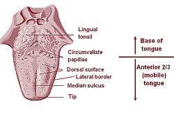
The lingual tonsils are two small mounds of lymphatic tissue located at the back of the base of the tongue, one on either side. They are composed of lymphatic tissue that functions to assist the immune system in the production of antibodies in response to invading pathogenic bacteria or viruses. If the tonsils are repeatedly swollen or infected over an extended period of time, they may need to be removed.
Function

Like other lymphatic tissues, the function of lingual tonsils is to prevent infections. These tonsils contain B and T lymphocytes which get activated when harmful bacteria and viruses come in contact with tonsils. B lymphocytes kill pathogens by producing antibodies against them, while T lymphocytes directly kill them by engulfing them.
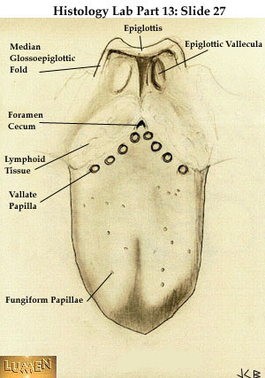
Histology
Lingual tonsils are covered externally by stratified squamous nonkeratinized epithelium. The epithelium invaginates inward to form a single crypt. Beneath the epithelium is a layer of lymphoid nodules containing lymphocytes. These tonsils are surrounded by thin capsule of connective tissue which separates them from adjacent structures. Mucous glands located at the root of tongue are drained through several ducts into the crypt of lingual tonsils. Secretions of these mucous glands keep the crypt clean and free of any debris. Therefore, the lingual tonsils are less prone to infection.
Blood supply
Lingual tonsils are located on posterior aspect of tongue which is supplied through:
- Lingual artery, branch of external carotid artery
- Tonsillar branch of facial artery
- Ascending pharyngeal branch of external carotid artery
Nerve supply
Lingual tonsils are innervated by tonsillar branches of glossopharyngeal nerve.That is located at the back of the tongue.
Pathology
An infection of the lingual tonsils causes many uncomfortable symptoms, including a sore throat and very painful swallowing. Swollen tonsils are often easily visible on the back of both sides of the tongue, a condition called tonsillitis. A fever may develop as the immune system tries to fight off the infection. A medical professional can visibly examine the tonsils to see if a bacterial culture should be made from a throat swab. Preliminary antibiotics may be prescribed until the results of the culture have returned from the laboratory. If no bacteria are revealed in the cultures, the antibiotics will be discontinued. If the infection is caused by a virus, the healthcare professional may be able to prescribe medication to relieve the painful sore throat and other uncomfortable symptoms. A solution of salt and water can be gargled to relieve some of the sore throat pain. The salt water also acts as an antiseptic rinse for the tonsil.
Extreme cases
In extreme cases, the inflamed lingual tonsil may grow large enough to block the airway, which can make breathing and speaking very difficult. Some people develop obstructive sleep apnea as the tonsils enlarge. A surgical procedure called coblation lingual tonsillectomy is used to remove the inflamed tonsils and restore the airway.
Surgery
During the tonsillectomy, the patient is put to sleep with general anesthesia. The surgeon removes the lingual tissue and seals the excision area. After the procedure, antibiotics and painkillers are prescribed for up to 10 days. A diet of soft and cold foods is recommended for at least the first three days after surgery.
Clinical
Lingual tonsillitis
Infection of lingual tonsils causes their inflammation, a condition called tonsillitis. Inflammation of these tonsils makes swallowing very difficult and painful. The other symptoms of tonsillitis are sore throat and fever.
Hypertrophy of lingual tonsils
Sometimes, the lingual tonsils become so enlarged that they obstruct the respiratory passage, leading to breathing difficulty. These hypertrophied tonsils are removed through tonsillectomy.
Lingual tonsils cancer
Squamous cell carcinoma of lingual tonsils occurs in people above 50 years of age.
Additional images
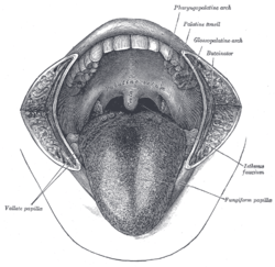 Lingual tonsils not labeled, but would be at very back of tongue
 Lymphatics of the tongue
 Lingual tonsil
 Lingual tonsil
 Lingual tonsils
References
This article incorporates text in the public domain from the 20th edition of Gray's Anatomy (1918)
External links
- Pictures at usc.edu(Registration required)
- Anatomy Atlases - Microscopic Anatomy, plate 09.163
- Histology image: 09802loa – Histology Learning System at Boston University
- MedEd at Loyola histo/HistoImages/hl6-27.jpg (labeled as 'lymphoid tissue')]
- Lingual Tonsil