Levator palpebrae superioris muscle
| Levator muscle of upper eyelid | |
|---|---|
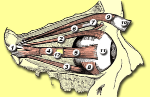 Rectus muscles: 2 = superior, 3 = inferior, 4 = medial, 5 = lateral Oblique muscles: 6 = superior, 8 = inferior Other muscle: 9 = levator palpebrae superioris Other structures: 1 = Annulus of Zinn, 7 = Trochlea, 10 = Superior tarsus, 11 = Sclera, 12 = Optic nerve | |
 Sagittal section of right orbital cavity. | |
| Details | |
| Origin | Sphenoid bone |
| Insertion | Tarsal plate, upper eyelid |
| Artery | Ophthalmic artery, superior ophthalmic vein |
| Nerve | Oculomotor nerve |
| Actions | Retracts / elevates eyelid |
| Antagonist | Orbicularis oculi muscle |
| Identifiers | |
| Latin | Musculus levator palpebrae superioris |
| TA | A15.2.07.020 |
| FMA | 49041 |
The levator palpebrae superioris (Latin for: elevating muscle of upper eyelid) is the muscle in the orbit that elevates the superior (upper) eyelid.
Structure
The levator palpebrae superioris originates on the lesser wing of the sphenoid bone, just above the optic foramen. It broadens and decreases in thickness (becomes thinner) and becomes the levator aponeurosis. This portion inserts on the skin of the upper eyelid, as well as the superior tarsal plate. It is a skeletal muscle. The superior tarsal muscle, a smooth muscle, is attached to the levator palpebrae superioris, and inserts on the superior tarsal plate as well.
Innervation
As with most of the muscles of the orbit, it is innervated by the superior division of the oculomotor nerve (Cranial Nerve III). It originates from a single central nucleus which provides innervation bilaterally to both LPS muscles. Due to this a single lesion in the central nucleus can cause bilateral ptosis. This is why, when one turns one's eye upward, the eyelid tends to rise with it.[1] An adjoining smooth muscle, the superior tarsal muscle, is sympathetically innervated and is occasionally considered to be part of the levator palpebrae superioris.
Function
The levator palpebrae superioris muscle elevates and retracts the upper eyelid.
Clinical signifiance
Damage to this muscle, or its innervation, can cause ptosis, the drooping of the eyelid. Ptosis can also be caused by damage to the adjoining superior tarsal muscle, or its sympathetic innervation. Such damage to the sympathetic supply occurs in Horner's syndrome, and presents as a partial ptosis.
Additional images
 The sinuses at the base of the skull.
 Nerves of the orbit. Seen from above.
 Dissection showing origins of right ocular muscles, and nerves entering by the superior orbital fissure.
 The right eye in sagittal section, showing the fascia bulbi (semidiagrammatic).
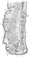 Sagittal section through the upper eyelid.
 Levator palpebrae superioris muscle
 Levator palpebrae superioris muscle
 Extrinsic eye muscle. Nerves of orbita. Deep dissection.
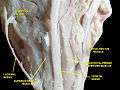 Extrinsic eye muscle. Nerves of orbita. Deep dissection.
See also
References
- ↑ "eye, human."Encyclopædia Britannica. 2008. Encyclopædia Britannica 2006 Ultimate Reference Suite DVD 5 Apr. 2008
External links
- -1979318192 at GPnotebook
- Anatomy figure: 29:01-01 at Human Anatomy Online, SUNY Downstate Medical Center
- lesson3 at The Anatomy Lesson by Wesley Norman (Georgetown University) (orbit2)