Introduction to viruses
| ||||||||||||
|
A single, fully functional virus particle outside its host cell Symmetry
| ||||||||||||
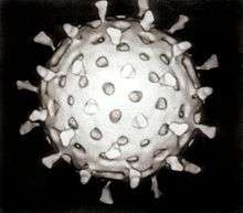
A virus is a biological agent that reproduces inside the cells of living hosts. When infected by a virus, a host cell is forced to produce many thousands of identical copies of the original virus at an extraordinary rate. Unlike most living things, viruses do not have cells that divide; new viruses are assembled in the infected host cell. But unlike still simpler infectious agents, viruses contain genes, which gives them the ability to mutate and evolve. Over 5,000 species of viruses have been discovered.[1]
The origins of viruses are unclear: some may have evolved from plasmids—pieces of DNA that can move between cells—while others may have evolved from bacteria. A virus consists of two or three parts: genes, made from either DNA or RNA, long molecules that carry genetic information; a protein coat that protects the genes; and in some viruses, an envelope of fat that surrounds the protein coat and is used, in combination with specific receptors, to enter a new host cell. Viruses vary in shape from the simple helical and icosahedral to more complex structures. Viruses range in size from 20 to 300 nanometres; it would take 30,000 to 750,000 of them, side by side, to stretch to 1 centimetre (0.39 in).
Viruses spread in many ways. Just as many viruses are very specific as to which host species or tissue they attack, each species of virus relies on a particular method for propagation. Plant viruses are often spread from plant to plant by insects and other organisms, known as vectors. Some viruses of animals, including humans, are spread by exposure to infected bodily fluids. Viruses such as influenza are spread through the air by droplets of moisture when people cough or sneeze. Viruses such as norovirus are transmitted by the faecal–oral route, which involves the contamination of hands, food and water. Rotavirus is often spread by direct contact with infected children. The human immunodeficiency virus, HIV, is transmitted by bodily fluids transferred during sex. Others, such as the Dengue virus, are spread by blood-sucking insects.
Viral infections can cause disease in humans, animals and even plants. However, they are usually eliminated by the immune system, conferring lifetime immunity to the host for that virus. Antibiotics have no effect on viruses, but antiviral drugs have been developed to treat life-threatening infections. Vaccines that produce lifelong immunity can prevent some viral infections.
Discovery

In 1884 the French microbiologist Charles Chamberland invented a filter, known today as the Chamberland filter or Chamberland–Pasteur filter, that has pores smaller than bacteria. Thus he could pass a solution containing bacteria through the filter and completely remove them from the solution.[2] In the early 1890s the Russian biologist Dmitri Ivanovsky used this filter to study what became known as the tobacco mosaic virus. His experiments showed that extracts from the crushed leaves of infected tobacco plants remain infectious after filtration.
At the same time several other scientists proved that, although these agents (later called viruses) were different from bacteria, they could still cause disease, and they were about one hundredth the size of bacteria. In 1899 the Dutch microbiologist Martinus Beijerinck observed that the agent multiplied only in dividing cells. Having failed to demonstrate its particulate nature he called it a "contagium vivum fluidum", a "soluble living germ".[3] In the early 20th century the English bacteriologist Frederick Twort discovered viruses that infect bacteria,[4] and the French-Canadian microbiologist Félix d'Herelle described viruses that, when added to bacteria growing on agar, would lead to the formation of whole areas of dead bacteria. Counting these dead areas allowed him to calculate the number of viruses in the suspension.[5]
With the invention of the electron microscope in 1931 by the German engineers Ernst Ruska and Max Knoll came the first images of viruses.[6] In 1935 American biochemist and virologist Wendell Meredith Stanley examined the tobacco mosaic virus and found it to be mostly made from protein.[7] A short time later, this virus was separated into protein and RNA parts.[8] A problem for early scientists was that they did not know how to grow viruses without using live animals. The breakthrough came in 1931, when the American pathologist Ernest William Goodpasture and Alice Miles Woodruff grew influenza and several other viruses in fertilised chickens' eggs.[9] Some viruses could not be grown in chickens' eggs, but this problem was solved in 1949 when John Franklin Enders, Thomas Huckle Weller and Frederick Chapman Robbins grew polio virus in cultures of living animal cells.[10] Over 5,000 species of virus have been discovered.[11]
Origins
Viruses co-exist with life wherever it occurs. They have probably existed since living cells first evolved. The origin of viruses remains unclear because they do not form fossils, so molecular techniques have been the most useful means of hypothesising how they arose. However, these techniques rely on the availability of ancient viral DNA or RNA but most of the viruses that have been preserved and stored in laboratories are less than 90 years old.[12][13] Molecular methods have only been successful in tracing the ancestry of viruses that evolved in the 20th century.[14] Three main theories speculate on the origins of viruses:[15][16]
- Regressive theory
- Viruses may have once been small cells that parasitised larger cells. Over time, genes not required by their parasitism were lost. The bacteria rickettsia and chlamydia are living cells that, like viruses, can reproduce only inside host cells. They lend credence to this theory, as their dependence on parasitism is likely to have caused the loss of genes that enabled them to survive outside a cell.[17]
- Cellular origin theory
- Some viruses may have evolved from bits of DNA or RNA that "escaped" from the genes of a larger organism. The escaped DNA could have come from plasmids—pieces of DNA that can move between cells—while others may have evolved from bacteria.[18]
- Coevolution theory
- Viruses may have evolved from complex molecules of protein and DNA at the same time as cells first appeared on earth and would have depended on cellular life for many millions of years.[19]
There are problems with all of these hypotheses: the regressive hypothesis does not explain why even the smallest of cellular parasites do not resemble viruses in any way. The escape hypothesis does not explain the structures of virus particles. The coevolution, or virus-first hypothesis, contravenes the definition viruses, in that they are dependent on host cells.[19] But viruses are recognised as ancient and to have origins that pre-date the divergence of life into the three domains.[20] This discovery has led modern virologists to reconsider and re-evaluate these three classical hypotheses.[20]
Structure

A virus particle, also known as a virion, consists of genes made from DNA or RNA which are surrounded by a protective coat of protein called a capsid.[21] The capsid is made of many smaller, identical protein molecules which are called capsomers. The arrangement of the capsomers can either be icosahedral (20-sided), helical or more complex. There is an inner shell around the DNA or RNA called the nucleocapsid, which is formed by proteins. Some viruses are surrounded by a bubble of lipid (fat) called an envelope.
Size
Viruses are among the smallest infectious agents, and most of them can only be seen by electron microscopy. Most viruses cannot be seen by light microscopy (in other words, they are sub-microscopic); their sizes range from 20 to 300 nm. They are so small that it would take 30,000 to 750,000 of them, side by side, to stretch to one cm.[21] By contrast bacterial sizes are typically around 1 micrometre (1000 nm) in diameter, and the cells of higher organisms a few tens of micrometres. Some viruses such as megaviruses and pandoraviruses are relatively large. At around 1 micrometer, these viruses, which infect amoebae, were discovered in 2003 and 2013. They are around a thousand times larger than influenza viruses and the discovery of these "giant" viruses astonished scientists.[22]
Genes
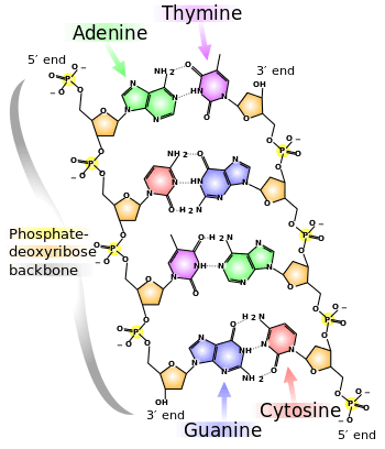
Genes are made from DNA (deoxyribonucleic acid) and, in many viruses, RNA (ribonucleic acid). The biological information contained in an organism is encoded in its DNA or RNA. Most organisms use DNA, but many viruses have RNA as their genetic material. The DNA or RNA of viruses consists of either a single strand or a double helix.[23]
Viruses reproduce rapidly because they have only a few genes compared to humans who have 20,000–25,000.[24] For example, influenza virus has only eight genes and rotavirus has eleven. These genes encode structural proteins that form the virus particle, or non-structural proteins, that are only found in cells infected by the virus.[25]
All cells, and many viruses, produce proteins that are enzymes called DNA polymerase and RNA polymerase which make new copies of DNA and RNA. A virus's polymerase enzymes are often much more efficient at making DNA and RNA than the host cell's.[26] However, RNA polymerase enzymes often make mistakes, and this is one of the reasons why RNA viruses often mutate to form new strains.[27]
In some species of RNA virus, the genes are not on a continuous molecule of RNA, but are separated. The influenza virus, for example, has eight separate genes made of RNA. When two different strains of influenza virus infect the same cell, these genes can mix and produce new strains of the virus in a process called reassortment.[28]
Protein synthesis

Proteins are essential to life. Cells produce new protein molecules from amino acid building blocks based on information coded in DNA. Each type of protein is a specialist that usually only performs one function, so if a cell needs to do something new, it must make a new protein. Viruses force the cell to make new proteins that the cell does not need, but are needed for the virus to reproduce. Protein synthesis consists of two major steps: transcription and translation.
Transcription is the process where information in DNA, called the genetic code, is used to produce RNA copies called messenger RNA (mRNA). These migrate through the cell and carry the code to ribosomes where it is used to make proteins. This is called translation because the protein's amino acid structure is determined by the mRNA's code. Information is hence translated from the language of nucleic acids to the language of amino acids.
Some nucleic acids of RNA viruses function directly as mRNA without further modification. For this reason, these viruses are called positive-sense RNA viruses.[29] In other RNA viruses, the RNA is a complementary copy of mRNA and these viruses rely on the cell's or their own enzyme to make mRNA. These are called negative-sense RNA viruses. In viruses made from DNA, the method of mRNA production is similar to that of the cell. The species of viruses called retroviruses behave completely differently: they have RNA, but inside the host cell a DNA copy of their RNA is made with the help of the enzyme reverse transcriptase. This DNA is then incorporated into the host's own DNA, and copied into mRNA by the cell's normal pathways.[30]
Life-cycle

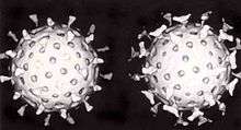
When a virus infects a cell, the virus forces it to make thousands more viruses. It does this by making the cell copy the virus's DNA or RNA, making viral proteins, which all assemble to form new virus particles.[31]
There are six basic, overlapping stages in the life cycle of viruses in living cells:[32]
- Attachment is the binding of the virus to specific molecules on the surface of the cell. This specificity restricts the virus to a very limited type of cell. For example, the human immunodeficiency virus (HIV) infects only human T cells, because its surface protein, gp120, can only react with CD4 and other molecules on the T cell's surface. Plant viruses can only attach to plant cells and cannot infect animals. This mechanism has evolved to favour those viruses that only infect cells in which they are capable of reproducing.
- Penetration follows attachment; viruses penetrate the host cell by endocytosis or by fusion with the cell.
- Uncoating happens inside the cell when the viral capsid is removed and destroyed by viral enzymes or host enzymes, thereby exposing the viral nucleic acid.
- Replication of virus particles is the stage where a cell uses viral messenger RNA in its protein synthesis systems to produce viral proteins. The RNA or DNA synthesis abilities of the cell produce the virus's DNA or RNA.
- Assembly takes place in the cell when the newly created viral proteins and nucleic acid combine to form hundreds of new virus particles.
- Release occurs when the new viruses escape or are released from the cell. Most viruses achieve this by making the cells burst, a process called lysis. Other viruses such as HIV are released more gently by a process called budding.
Effects on the host cell
The range of structural and biochemical effects that viruses have on the host cell is extensive.[33] These are called cytopathic effects.[34] Most virus infections eventually result in the death of the host cell. The causes of death include cell lysis (bursting), alterations to the cell's surface membrane and apoptosis (cell "suicide").[35] Often cell death is caused by cessation of its normal activity due to proteins produced by the virus, not all of which are components of the virus particle.[36]
Some viruses cause no apparent changes to the infected cell. Cells in which the virus is latent and inactive show few signs of infection and often function normally.[37] This causes persistent infections and the virus is often dormant for many months or years. This is often the case with herpes viruses.[38][39]
Some viruses, such as Epstein-Barr virus, often cause cells to proliferate without causing malignancy;[40] but some other viruses, such as papillomavirus, are an established cause of cancer.[41] When a cell's DNA is damaged by a virus, and if the cell cannot repair itself, this often triggers apoptosis. One of the results of apoptosis is destruction of the damaged DNA by the cell itself. Some viruses have mechanisms to limit apoptosis so that the host cell does not die before progeny viruses have been produced; HIV, for example, does this.[35]
Viruses and diseases

Common human diseases caused by viruses include the common cold, the flu, chickenpox and cold sores. Serious diseases such as Ebola and AIDS are also caused by viruses. Many viruses cause little or no disease and are said to be "benign". The more harmful viruses are described as virulent. Viruses cause different diseases depending on the types of cell that they infect. Some viruses can cause lifelong or chronic infections where the viruses continue to reproduce in the body despite the host's defence mechanisms.[42] This is common in hepatitis B virus and hepatitis C virus infections. People chronically infected with a virus are known as carriers. They serve as important reservoirs of the virus. If there is a high proportion of carriers in a given population, a disease is said to be endemic.[43]
There are many ways in which viruses spread from host to host but each species of virus uses only one or two. Many viruses that infect plants are carried by organisms; such organisms are called vectors. Some viruses that infect animals, including humans, are also spread by vectors, usually blood-sucking insects. However, direct transmission is more common. Some virus infections, such as norovirus and rotavirus, are spread by contaminated food and water, hands and communal objects and by intimate contact with another infected person,[44] while others are airborne (influenza virus).[45] Viruses such as HIV, hepatitis B and hepatitis C are often transmitted by unprotected sex[46] or contaminated hypodermic needles.[47] It is important to know how each different kind of virus is spread to prevent infections and epidemics.[48]
Diseases of plants

There are many types of plant virus, but often they only cause a loss of yield, and it is not economically viable to try to control them. Plant viruses are often spread from plant to plant by organisms (vectors). These are normally insects, but some fungi, nematode worms and single-celled organisms have been shown to be vectors. When control of plant virus infections is considered economical (perennial fruits, for example) efforts are concentrated on killing the vectors and removing alternate hosts such as weeds.[49] Plant viruses are harmless to humans and other animals because they can only reproduce in living plant cells.[50]
Bacteriophages
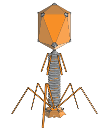
Bacteriophages are viruses that infect bacteria and archaea. The International Committee on Taxonomy of Viruses officially recognises 28 genera of bacteriophages that belong to 11 families.[51] They are important in marine ecology: as the infected bacteria burst, carbon compounds are released back into the environment, which stimulates fresh organic growth. Bacteriophages are useful in scientific research because they are harmless to humans and can be studied easily. These viruses can be a problem in industries that produce food and drugs by fermentation and depend on healthy bacteria. Some bacterial infections are becoming difficult to control with antibiotics, so there is a growing interest in the use of bacteriophages to treat infections in humans.[52]
Host resistance
Innate immunity of animals
Animals, including humans, have many natural defences against viruses. Some are non-specific and protect against many viruses regardless of the type. This innate immunity is not improved by repeated exposure to viruses and does not retain a "memory" of the infection. The skin of animals, particularly its surface, which is made from dead cells, prevents many types of viruses from infecting the host. The acidity of the contents of the stomach destroys many viruses that have been swallowed. When a virus overcomes these barriers and enters the host, other innate defences prevent the spread of infection in the body. A special hormone called interferon is produced by the body when viruses are present, and this stops the viruses from reproducing by killing the infected cell and its close neighbours. Inside cells, there are enzymes that destroy the RNA of viruses. This is called RNA interference. Some blood cells engulf and destroy other virus infected cells.[53]
Adaptive immunity of animals
Specific immunity to viruses develops over time and white blood cells called lymphocytes play a central role. Lymphocytes retain a "memory" of virus infections and produce many special molecules called antibodies. These antibodies attach to viruses and stop the virus from infecting cells. Antibodies are highly selective and attack only one type of virus. The body makes many different antibodies, especially during the initial infection; however, after the infection subsides, some antibodies remain and continue to be produced, often giving the host lifelong immunity to the virus.[54]
Plant resistance
Plants have elaborate and effective defence mechanisms against viruses. One of the most effective is the presence of so-called resistance (R) genes. Each R gene confers resistance to a particular virus by triggering localised areas of cell death around the infected cell, which can often be seen with the unaided eye as large spots. This stops the infection from spreading.[55] RNA interference is also an effective defence in plants.[56] When they are infected, plants often produce natural disinfectants which destroy viruses, such as salicylic acid, nitric oxide and reactive oxygen molecules.[57]
Resistance to bacteriophages
The major way bacteria defend themselves from bacteriophages is by producing enzymes which destroy foreign DNA. These enzymes, called restriction endonucleases, cut up the viral DNA that bacteriophages inject into bacterial cells.
Prevention and treatment of viral disease in humans and other animals
Vaccines
Vaccination is a way of preventing diseases caused by viruses. Vaccines simulate a natural infection and its associated immune response, but do not cause the disease. Their use has resulted in the eradication of smallpox and a dramatic decline in illness and death caused by infections such as polio, measles, mumps and rubella.[58] Vaccines are available to prevent over thirteen viral infections of humans[59] and more are used to prevent viral infections of animals.[60] Vaccines may consist of either live or killed viruses.[61] Live vaccines contain weakened forms of the virus, but these vaccines can be dangerous when given to people with weak immunity. In these people, the weakened virus can cause the original disease.[62] Biotechnology and genetic engineering techniques are used to produce "designer" vaccines that only have the capsid proteins of the virus. Hepatitis B vaccine is an example of this type of vaccine.[63] These vaccines are safer because they can never cause the disease.[64]
Antiviral drugs
Over the past 20 years, the development of antiviral drugs has increased rapidly, mainly driven by the AIDS pandemic. Antiviral drugs are often nucleoside analogues, which are molecules very similar, but not identical to DNA building blocks. When the replication of virus DNA begins, some of these fake building blocks are incorporated. As soon as that happens, replication stops prematurely—the fake building blocks lack the essential features that allow the addition of further building blocks. Thus, DNA production is halted, and the virus can no longer reproduce.[65] Examples of nucleoside analogues are aciclovir for herpes virus infections and lamivudine for HIV and hepatitis B virus infections. Aciclovir is one of the oldest and most frequently prescribed antiviral drugs.[66]

Other antiviral drugs target different stages of the viral life cycle. HIV is dependent on an enzyme called the HIV-1 protease for the virus to become infectious. There is a class of drugs called protease inhibitors, which bind to this enzyme and stop it from functioning.[67]
Hepatitis C is caused by an RNA virus. In 80% of people infected, the disease becomes chronic, and they remain infectious for the rest of their lives unless they are treated. There is an effective treatment that uses the nucleoside analogue drug ribavirin combined with interferon.[68] Treatments for chronic carriers of the hepatitis B virus by a similar strategy using lamivudine and other anti-viral drugs have been developed.[69] In both diseases, the drugs stop the virus from reproducing and the interferon kills any remaining infected cells.
HIV infections are usually treated with a combination of antiviral drugs, each targeting a different stage in the virus's life-cycle. There are drugs that prevent the virus from attaching to cells, others that are nucleoside analogues and some poison the virus's enzymes that it needs to reproduce.[67] The success of these drugs is proof of the importance of knowing how viruses reproduce.
Role in ecology
Viruses are the most abundant biological entity in aquatic environments[70]—there are about one million of them in a teaspoon of seawater[71]—and they are essential to the regulation of saltwater and freshwater ecosystems.[72] Most of these viruses are bacteriophages, which are harmless to plants and animals. They infect and destroy the bacteria in aquatic microbial communities and this is the most important mechanism of recycling carbon in the marine environment. The organic molecules released from the bacterial cells by the viruses stimulate fresh bacterial and algal growth.[73]
Microorganisms constitute more than 90% of the biomass in the sea. It is estimated that viruses kill approximately 20% of this biomass each day and that there are fifteen times as many viruses in the oceans as there are bacteria and archaea. Viruses are mainly responsible for the rapid destruction of harmful algal blooms,[74] which often kill other marine life.[75] The number of viruses in the oceans decreases further offshore and deeper into the water, where there are fewer host organisms.[76]
Their effects are far-reaching; by increasing the amount of respiration in the oceans, viruses are indirectly responsible for reducing the amount of carbon dioxide in the atmosphere by approximately 3 gigatonnes of carbon per year.[76]
Marine mammals are also susceptible to viral infections. In 1988 and 2002, thousands of harbour seals were killed in Europe by phocine distemper virus.[77] Many other viruses, including caliciviruses, herpesviruses, adenoviruses and parvoviruses, circulate in marine mammal populations.[76]
See also
References
Notes
- ↑ Leppard, Keith; Nigel Dimmock; Easton, Andrew (2007). Introduction to Modern Virology. Blackwell Publishing Limited. p. 4. ISBN 1-4051-3645-6.
- ↑ Shors pp. 76–77
- ↑ Topley and Wilson p. 3
- ↑ Shors p. 589
- ↑ D'herelle, F (2007). "On an invisible microbe antagonistic toward dysenteric bacilli: brief note by Mr. F. D'Herelle, presented by Mr. Roux. 1917". Research in microbiology. 158 (7): 553–4. doi:10.1016/j.resmic.2007.07.005. PMID 17855060.
- ↑ From Nobel Lectures, Physics 1981–1990, (1993) Editor-in-Charge Tore Frängsmyr, Editor Gösta Ekspång, World Scientific Publishing Co., Singapore
- ↑ Stanley, W.M.; Loring, H.S. (1936). "The isolation of crystalline tobacco mosaic virus protein from diseased tomato plants". Science. 83 (2143): 85. Bibcode:1936Sci....83...85S. doi:10.1126/science.83.2143.85. PMID 17756690.
- ↑ Stanley, W.M.; Lauffer, M.A. (1939). "Disintegration of tobacco mosaic virus in urea solutions". Science. 89 (2311): 345–347. Bibcode:1939Sci....89..345S. doi:10.1126/science.89.2311.345. PMID 17788438.
- ↑ Goodpasture, E.W.; Woodruff, A.M.; Buddingh, G.J. (1931). "The cultivation of vaccine and other viruses in the chorioallantoic membrane of chick embryos". Science. 74 (1919): 371–372. Bibcode:1931Sci....74..371G. doi:10.1126/science.74.1919.371. PMID 17810781.
- ↑ Rosen, F.S. (2004). "Isolation of poliovirus—John Enders and the Nobel Prize". New England Journal of Medicine. 351 (15): 1481–83. doi:10.1056/NEJMp048202. PMID 15470207.
- ↑ Dimmock, N.J; Easton, Andrew J; Leppard, Keith (2007) Introduction to Modern Virology sixth edition, Blackwell Publishing, ISBN 1-4051-3645-6. p. 49
- ↑ Shors. p. 16
- ↑ Topley and Wilson pp. 18–19
- ↑ Liu, Y; Nickle, DC; Shriner, D; Jensen, MA; Learn Jr, GH; Mittler, JE; Mullins, JI (2004). "Molecular clock-like evolution of human immunodeficiency virus type 1.". Virology. 329 (1): 101–8. doi:10.1016/j.virol.2004.08.014. PMID 15476878.
- ↑ Shors pp. 14–16
- ↑ Topley and Wilson pp.11–21
- ↑ Topley and Wilson p. 11
- ↑ Topley and Wilson pp. 11–12
- 1 2 Wessner D. R. (2010). "The Origins of Viruses". Nature Education. 3 (9): 37.
- 1 2 Mahy WJ & Van Regenmortel MHV (eds). Desk Encyclopedia of General Virology. Oxford: Academic Press; 2009. ISBN 0-12-375146-2. p. 28.
- 1 2 Topley and Wilson pp. 33–55
- ↑ Zimmer, Carl (18 July 2013). "Changing View on Viruses: Not So Small After All". New York Times. Retrieved 20 December 2014.
- ↑ Shors pp. 54–61
- ↑ International Human, Genome Sequencing Consortium (2004). "Finishing the euchromatic sequence of the human genome". Nature. 431 (7011): 931–945. Bibcode:2004Natur.431..931H. doi:10.1038/nature03001. PMID 15496913.
- ↑ Shors p. 73
- ↑ Shors pp. 32–34
- ↑ Shors p. 510
- ↑ Shors p. 327
- ↑ Topley and Wilson pp. 75–82
- ↑ Shors pp. 248–250
- ↑ Shors pp. 11–12
- ↑ Shors pp. 47–67
- ↑ Collier pp. 115–146
- ↑ Collier p. 115
- 1 2 Roulston A, Marcellus RC, Branton PE; Marcellus; Branton (1999). "Viruses and apoptosis". Annu. Rev. Microbiol. 53 (1): 577–628. doi:10.1146/annurev.micro.53.1.577. PMID 10547702. Retrieved 2014-12-20.
- ↑ Alwine JC (2008). "Modulation of host cell stress responses by human cytomegalovirus". Curr. Top. Microbiol. Immunol. Current Topics in Microbiology and Immunology. 325: 263–79. doi:10.1007/978-3-540-77349-8_15. ISBN 978-3-540-77348-1. PMID 18637511.
- ↑ Sinclair J (March 2008). "Human cytomegalovirus: Latency and reactivation in the myeloid lineage". J. Clin. Virol. 41 (3): 180–5. doi:10.1016/j.jcv.2007.11.014. PMID 18164651. Retrieved 2014-12-20.
- ↑ Jordan MC, Jordan GW, Stevens JG, Miller G; Jordan; Stevens; Miller (June 1984). "Latent herpesviruses of humans". Ann. Intern. Med. 100 (6): 866–80. doi:10.7326/0003-4819-100-6-866. PMID 6326635.
- ↑ Sissons JG, Bain M, Wills MR; Bain; Wills (February 2002). "Latency and reactivation of human cytomegalovirus". J. Infect. 44 (2): 73–7. doi:10.1053/jinf.2001.0948. PMID 12076064. Retrieved 2014-12-20.
- ↑ Barozzi P, Potenza L, Riva G, Vallerini D, Quadrelli C, Bosco R, Forghieri F, Torelli G, Luppi M; Potenza; Riva; Vallerini; Quadrelli; Bosco; Forghieri; Torelli; Luppi (December 2007). "B cells and herpesviruses: a model of lymphoproliferation". Autoimmun Rev. 7 (2): 132–6. doi:10.1016/j.autrev.2007.02.018. PMID 18035323. Retrieved 2014-12-20.
- ↑ Subramanya D, Grivas PD; Grivas (November 2008). "HPV and cervical cancer: updates on an established relationship". Postgrad Med. 120 (4): 7–13. doi:10.3810/pgm.2008.11.1928. PMID 19020360.
- ↑ Shors p. 483
- ↑ Topley and Wilson p. 766
- ↑ Shors p. 118
- ↑ Shors p.117
- ↑ Shors p. 119
- ↑ Shors p.123
- ↑ Shors pp. 16–19
- ↑ Shors p. 584
- ↑ Shors pp. 562–587
- ↑ Fauquet, CM (2009). Desk Encyclopedia of General Virology. Boston: Academic Press. p. 82. ISBN 0-12-375146-2.
- ↑ Shors pp. 588–604
- ↑ Shors pp. 146–158
- ↑ Shors pp.158–168
- ↑ Dinesh-Kumar, S.P.; Tham, Hong; -1#Wai-, Baker (2000). "Structure—function analysis of the tobacco mosaic virus resistance gene N". PNAS. 97 (26): 14789–94. Bibcode:2000PNAS...9714789D. doi:10.1073/pnas.97.26.14789. PMC 18997
 . PMID 11121079.
 - ↑ Shors pp. 573–576; Ding, S. W.; Voinnet, O. (2007). "Antiviral Immunity Directed by Small RNAs". Cell. 130 (3): 413–426. doi:10.1016/j.cell.2007.07.039. PMC 2703654. PMID 17693253. PMID 17693253
- ↑ Soosaar, J.L.; Burch-Smith, T.M.; Dinesh-Kumar, S.P. (2005). "Mechanisms of plant resistance to viruses". Nature Reviews Microbiology. 3 (10): 789–98. doi:10.1038/nrmicro1239. PMID 16132037.
- ↑ Shors pp. 171–185
- ↑ Shors p. 183
- ↑ Pastoret, P.P.; Schudel, A.A.; Lombard, M. (2007). "Conclusions—future trends in veterinary vaccinology". Rev. Off. Int. Epizoot. 26 (2): 489–94, 495–501, 503–9. PMID 17892169.
- ↑ Shors p. 172
- ↑ Thomssen, R. (1975). "Live attenuated versus killed virus vaccines". Monographs in allergy. 9: 155–76. PMID 1090805.
- ↑ Shors p. 174
- ↑ Shors p. 180
- ↑ Shors p. 427
- ↑ Shors p. 426
- 1 2 Shors p. 463
- ↑ Witthoft, T.; Moller, B.; Wiedmann, K.H.; Mauss, S.; Link, R.; Lohmeyer, J.; Lafrenz, M.; Gelbmann, C.M.; Huppe, D.; et al. (2007). "Safety, tolerability and efficacy of peginterferon alpha-2a and ribavirin in chronic hepatitis C in clinical practice: The German Open Safety Trial". J Viral Hepat. 14 (11): 788–796. doi:10.1111/j.1365-2893.2007.00871.x. PMC 2156112. PMID 17927615.
- ↑ Paul N, Han SH; Han (June 2011). "Combination Therapy for Chronic Hepatitis B: Current Indications". Current Hepatitis Reports. 10 (2): 98–105. doi:10.1007/s11901-011-0095-1. PMC 3085106. PMID 21654909.
- ↑ Koonin EV, Senkevich TG, Dolja VV; Senkevich; Dolja (2006). "The ancient Virus World and evolution of cells". Biology Direct. 1 (1): 29. doi:10.1186/1745-6150-1-29. PMC 1594570. PMID 16984643.
- ↑ Shors p. 4
- ↑ Shors p. 5
- ↑ Shors p. 593
- ↑ Suttle CA (September 2005). "Viruses in the sea". Nature. 437 (7057): 356–61. Bibcode:2005Natur.437..356S. doi:10.1038/nature04160. PMID 16163346.
- ↑ "Harmful Algal Blooms: Red Tide: Home | CDC HSB". www.cdc.gov. Retrieved 23 August 2009.
- 1 2 3 Suttle CA (October 2007). "Marine viruses—major players in the global ecosystem". Nature Reviews Microbiology. 5 (10): 801–12. doi:10.1038/nrmicro1750. PMID 17853907.
- ↑ Hall, A; Jepson, P; Goodman, S; Harkonen, T (2006). "Phocine distemper virus in the North and European Seas – Data and models, nature and nurture". Biological Conservation. 131 (2): 221–229. doi:10.1016/j.biocon.2006.04.008.
Bibliography
- Collier, Leslie; Balows, Albert; Sussman Max (1998) Topley and Wilson's Microbiology and Microbial Infections ninth edition, Volume 1, Virology, volume editors: Mahy, Brian and Collier, Leslie. Arnold. ISBN 0-340-66316-2
- Shors, Teri (2008). Understanding Viruses. Jones and Bartlett Publishers. ISBN 0-7637-2932-9
External links
- Virus Pathogen Resource – Genomic and other research data about human pathogenic viruses
- Influenza Research Database – Genomic and other research data about influenza viruses
| Library resources about Virus |