Intraparietal sulcus
| Intraparietal sulcus | |
|---|---|
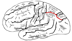 Lateral surface of left cerebral hemisphere, viewed from the side. (Intraparietal sulcus visible at upper right, running horizontally.) | |
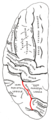
 Right cerebral hemisphere, viewed from the side. The region colored in blue is parietal lobe of the human brain. Intraparietal sulcus runs horizontally at the middle of the parietal lobe. | |
| Details | |
| Part of | Parietal lobe |
| Identifiers | |
| Latin | sulcus intraparietalis |
| Acronym(s) | IPS |
| NeuroNames | hier-79 |
| NeuroLex ID | Intraparietal sulcus |
| TA | A14.1.09.127 |
| FMA | 83772 |
The intraparietal sulcus (IPS) is located on the lateral surface of the parietal lobe, and consists of an oblique and a horizontal portion. The IPS contains a series of functionally distinct subregions that have been intensively investigated using both single cell neurophysiology in primates[1][2] and human functional neuroimaging.[3] Its principal functions are related to perceptual-motor coordination (e.g., directing eye movements and reaching) and visual attention, which allows for visually-guided pointing, grasping, and object manipulation that can produce a desired effect.
The IPS is also thought to play a role in other functions, including processing symbolic numerical information,[4] visuospatial working memory[5] and interpreting the intent of others.[6]
Function
Five regions of the intraparietal sulcus (IPS): anterior, lateral, ventral, caudal, and medial
- LIP & VIP: involved in visual attention and saccadic eye movements
- VIP & MIP: visual control of reaching and pointing
- AIP: visual control of grasping and manipulating hand movements
- CIP: perception of depth from stereopsis
All of these areas have projections to the frontal lobe for executive control.
Activity in the intraparietal sulcus has also been associated with the learning of sequences of finger movements.[7]
The task-positive network includes the intraparietal sulcus in each hemisphere;[8] it is one of two sensory orienting systems in the human brain.
Understanding numbers
Behavioral studies suggest that the IPS is associated with impairments of basic numerical magnitude processing and that there is a pattern of structural and functional alternations in the IPS and in the PFC in dyscalculia.[9] Children with developmental dyscalculia were found to have less gray matter in the left IPS.[10]
Studies have shown that electrical activity in a particular group of nerve cells in the intraparietal sulcus spiked when, and only when, volunteers were performing calculations. Outside experimental settings it was also found that when a patient mentioned a number — or even a quantitative reference, such as “some more,” “many” or “bigger than the other one” — there was a spike of electrical activity in the same nerve-cell population of the intraparietal sulcus that was activated when the patient was doing calculations under experimental conditions.[11]
Additional images
 Lateral surface of left cerebral hemisphere, viewed from above.
 Left cerebral hemisphere, viewed from the back. (Intraparietal sulcus visible at top center)
_Intraparietal_sulcus.webm.jpg) Human brain dissection video (53 sec). Demonstrating position of the intraparietal sulcus of the left cerebral hemisphere.
Human brain dissection video (53 sec). Demonstrating position of the intraparietal sulcus of the left cerebral hemisphere.
References
- ↑ Colby C.E.; Goldberg M.E. (1999). "Space and attention in parietal cortex". Annual Review of Neuroscience. 22: 319–349. doi:10.1146/annurev.neuro.22.1.319. PMID 10202542.
- ↑ Andersen R.A. (1989). "Visual and eye movement functions of the posterior parietal cortex". Annual Review of Neuroscience. 12: 377–403. doi:10.1146/annurev.ne.12.030189.002113. PMID 2648954.
- ↑ Culham, J.C.; Nancy G. Kanwisher (April 2001). "Neuroimaging of cognitive functions in human parietal cortex". Current Opinion in Neurobiology. 11 (2): 157–163. doi:10.1016/S0959-4388(00)00191-4.
- ↑ Cantlon J, Brannon E, Carter E, Pelphrey K (2006). "Functional imaging of numerical processing in adults and 4-y-old children.". PLoS Biol. 4 (5): e125. doi:10.1371/journal.pbio.0040125. PMC 1431577
 . PMID 16594732. link
 - ↑ Todd JJ, Marois R (2004). "Capacity limit of visual short-term memory in human posterior parietal cortex". Nature. 428 (6984): 751–754. doi:10.1038/nature02466. PMID 15085133.
- ↑ Grafton, Hamilton (2006). "Dartmouth Study Finds How The Brain Interprets The Intent Of Others.". Science Daily.
- ↑ Sakai, K.; Ramnani, N.; Passingham, R. E. (2002). "Learning of sequences of finger movements and timing: Frontal lobe and action-oriented representation". Journal of Neurophysiology. 88 (4): 2035–2046. PMID 12364526.
- ↑ Fox, M.D., Corbetta, M., Snyder, A.Z., Vincent, J.L., & Raichle, M.E. (2006). Spontaneous neuronal activity distinguishes human dorsal and ventral attention systems. Proceedings of the National Academy of Sciences, 103, 10046-10051.
- ↑ Ansari D.; Karmiloff-Smith A. (2002). "Atypical trajectories of number development: a neuroconstructivist perspective". Trends in Cognitive Science. 6 (12): 511–516. doi:10.1016/S1364-6613(02)02040-5. PMID 12475711.
- ↑ Kucian K, et al. (2006). "Impaired neural networks for approximate calculation in dyscalculic children: a functional MRI study". Behavior and Brain Function. 2: 31. doi:10.1186/1744-9081-2-31. PMC 1574332. PMID 16953876.
- ↑ Dastjerdi, M.; Ozker, M.; Foster, B. L.; Rangarajan, V.; Parvizi, J. (2013). "Numerical processing in the human parietal cortex during experimental and natural conditions". Nature Communications. 4. doi:10.1038/ncomms3528.
External links
| Wikimedia Commons has media related to Intraparietal sulcus. |