Interferometric scattering microscopy
Interferometric scattering microscopy (iSCAT) is an optical microscopy technique that relies on collecting light scattered by an object together with a reference light field, provided by the reflection at an interface. In the presence of the scattering object, the total intensity of light returning in the direction of the incident beam is different than in the absence of that object due to interference of the scattered and reflected light. Thus, the object appears as a small dip on top of a large background.[1]
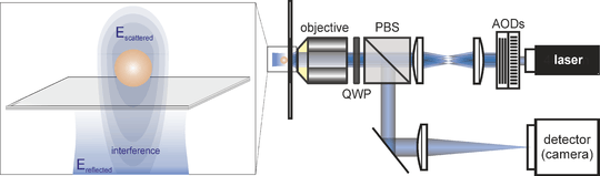
An iSCAT microscope requires illumination of the sample and collection of reflected and scattered light, which interfere at the detector. The concept is different compared to dark field microscopy, in the sense that iSCAT doesn't reject the reflected (background) light. The signal has therefore three main components: (1) reflected light, which dominates the iSCAT image and which is reduced to a minimum in dark field microscopy; (2) scattered light, which is negligible in iSCAT but the main signal in a dark field image; (3) interference term, which modulates the signal originating from reflection proportional to the scattering amplitude. iSCAT signal scales linearly with the scattering amplitude and thus particle's volume (it is V^2 in case of dark field scattering detection). The basic design of an interferometric detection has been explored for decades in interference reflection microscopy and differential interference contrast microscopy. The major difference between those techniques and iSCAT is that the latter uses a coherent light source, which increases the interferometric contrast.[2]
Biological applications:
- label-free virus tracking[3]
- GM1 tracking[4]
- molecular motors tracking[5]
- label-free imaging of lipid vesicles,[6] lipid nanodomains,[7] or myosin 5[8]

References
- ↑ Lindfors, K., Kalkbrenner, T., Stoller, P. & Sandoghdar, V. Detection and Spectroscopy of Gold Nanoparticles Using Supercontinuum White Light Confocal Microscopy. Phys.Rev.Lett. (2004)
- ↑ Ortega-Arroyo, J. & Kukura, P. Interferometric Scattering Microscopy (iSCAT): New Frontiers in Ultrafast and Ultrasensitive Optical Microscopy. Phys.Chem.Chem.Phys. (2012)
- ↑ Kukura, P. et al. High-Speed Nanoscopic Tracking of the Position and Orientation of a Single Virus. Nat.Methods (2009)
- ↑ Spillane, K. M. et al. High-speed single-particle tracking of GM1 in model membranes reveals anomalous diffusion due to interleaflet coupling and molecular pinning. Nano Lett (2014)
- ↑ Andrecka, J. et al. Structural dynamics of myosin 5 during processive motion revealed by interferometric scattering microscopy. eLife (2015)
- ↑ Andrecka, J., Spillane, K. M., Ortega-Arroyo, J. & Kukura, P. Direct observation and control of supported lipid bilayer formation with interferometric scattering microscopy. ACS nano (2013)
- ↑ de Wit, G., Danial, J. S., Kukura, P. & Wallace, M. I. Dynamic label-free imaging of lipid nanodomains. PNAS (2015)
- ↑ Ortega Arroyo, J. et al. Label-free, all-optical detection, imaging, and tracking of a single protein. Nano Lett. (2014)