Intercostal nerves
| Intercostal nerves | |
|---|---|
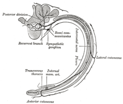 Diagram of the course and branches of a typical intercostal nerve. | |
 Intercostal nerves, the superficial muscles having been removed. | |
| Details | |
| From | thoracic nerves (T1-T11) |
| Innervates | intercostal muscle |
| Identifiers | |
| Latin | nervi intercostales |
| MeSH | A08.800.800.720.800.350 |
| TA | A14.2.04.006 |
| FMA | 75467 |
The intercostal nerves are part of the somatic nervous system, and arise from the anterior rami of the thoracic spinal nerves from T1 to T11. The intercostal nerves are distributed chiefly to the thoracic pleura and abdominal peritoneum and differ from the anterior rami of the other spinal nerves in that each pursues an independent course without plexus formation.
The first two nerves supply fibers to the upper limb in addition to their thoracic branches; the next four are limited in their distribution to the walls of the thorax; the lower five supply the walls of the thorax and abdomen. The 7th intercostal nerve terminates at the xyphoid process, at the lower end of the sternum. The 10th intercostal nerve terminates at the navel. The twelfth (subcostal) thoracic is distributed to the abdominal wall and groin.
Unlike the nerves from the autonomic nervous system that innervate the visceral pleura of the thoracic cavity, the intercostal nerves arise from the somatic nervous system. This enables them to control the contraction of muscles, as well as provide specific sensory information regarding the skin and parietal pleura. This explains why damage to the internal wall of the thoracic cavity can be felt as a sharp pain localized in the injured region. Damage to the visceral pleura is experienced as an un-localized ache.
The 1st Thoracic Nerve
The anterior division of the first thoracic nerve divides into two branches: one, the larger, leaves the thorax in front of the neck of the first rib, and enters the brachial plexus; the other and smaller branch, the first intercostal nerve, runs along the first intercostal space, and ends on the front of the chest as the first anterior cutaneous branch of the thorax.
Occasionally this anterior cutaneous branch is missing.
The first intercostal nerve rarely gives off a lateral cutaneous branch; but sometimes sends a small branch to communicate with the intercostobrachial.
From the second thoracic nerve it frequently receives a connecting twig, which ascends over the neck of the second rib. This nerve was first described by Kuntz in 1927. There is considerable anatomic variation, but Kuntz nerve may be present in 40-80% of the population.[1][2]
The Upper Thoracic Nerves: 2nd-6th
The anterior divisions of the second, third, fourth, fifth, and sixth thoracic nerves, and the small branch from the first thoracic, are confined to the walls of the thorax, and are named thoracic intercostal nerves.
They pass forward in the intercostal spaces below the intercostal vessels. At the back of the chest they lie between the pleura and the posterior intercostal membranes, but soon pierce the latter and run between the two planes of Intercostal muscles as far as the middle of the rib.
They then enter the substance of the Intercostales interni, and, running amidst their fibers as far as the costal cartilages, they gain the inner surfaces of the muscles and lie between them and the pleura.
Near the sternum, they cross in front of the internal mammary artery and Transversus thoracis muscle, pierce the Intercostales interni, the anterior intercostal membranes, and Pectoralis major, and supply the integument of the front of the thorax and over the mamma, forming the anterior cutaneous branches of the thorax; the branch from the second nerve unites with the anterior supraclavicular nerves of the cervical plexus.
The fourth intercostal nerve is enervated by cutaneous slowly-adapting and rapidly-adapting mechanoreceptors, especially by ones densly-packed under the areola; enervation subsequently triggers oxytocin release, which, when in the peripheral bloodstream, causes myoepithelial cell contraction and lactation: this is an example of a non-nerve-innervation muscular reflex.
Branches
Numerous slender muscular filaments supply the Intercostales, the Subcostales, the Levatores costarum, the Serratus posterior superior, and the Transversus thoracis. At the front of the thorax some of these branches cross the costal cartilages from one intercostal space to another.
- Lateral cutaneous branches (rami cutanei laterales) are derived from the intercostal nerves, about midway between the vertebræ and sternum; they pierce the Intercostales externi and Serratus anterior, and divide into anterior and posterior branches.
- The anterior branches run forward to the side and the forepart of the chest and skin, fourth nerve anterior branches supplying the areola and the mamma; those of the fifth and sixth nerves supply the upper digitations of the Obliquus externus abdominis.
- The posterior branches run backward, and supply the skin over the scapula and Latissimus dorsi.
The lateral cutaneous branch of the second intercostal nerve does not divide, like the others, into an anterior and a posterior branch; it is named the intercostobrachial nerve.
The Lower Thoracic Nerves: 7th-11th
The Lower Thoracic Nerves: 12th
Anterior division
- See subcostal nerve
Lateral cutaneous branch
The lateral cutaneous branch of the last thoracic nerve is large, and undivided.
It perforates the internal and the external oblique muscles, descends over the iliac crest in front of the lateral cutaneous branch of the iliohypogastric nerve, and is distributed to the skin of the front part of the gluteal muscles, some of its filaments extending as low as the greater trochanter of the femur.
Additional images
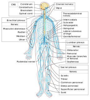 Nervous system
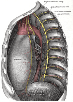 Intercostal spaces, viewed from the left
 Brachial plexus
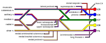 Brachial plexus with courses of spinal nerves shown
See also
- Peripheral nervous system
- Intercostales externi muscle
- Intercostales interni muscle
- Intercostal nerve block
References
This article incorporates text in the public domain from the 20th edition of Gray's Anatomy (1918)
- ↑ Ramsaroop L, Partab P, Singh B, Satyapal KS. Thoracic origin of a sympathetic supply to the upper limb: the ‘nerve of Kuntz’ revisited. J Anat. 2001;199:675Y682
- ↑ Marhold F, Izay B, Zacherl J, Tschabitscher M, Neumayer C. Thoracoscopic and anatomic landmarks of Kuntz’s nerve: implications for sympathetic surgery. Ann Thorac Surg. 2008;86:1653Y1658.
External links
- thoraxlesson5 at The Anatomy Lesson by Wesley Norman (Georgetown University) (paravertebralregion)
- Atlas image: abdo_wall53 at the University of Michigan Health System - "Abdominal Wall, Dissection, Lateral View"