Inferior frontal gyrus
| Inferior frontal gyrus | |
|---|---|
 Inferior frontal gyrus of the human brain, gyrus frontalis inferior. | |
 Lateral surface of left cerebral hemisphere, viewed from the side. | |
| Details | |
| Part of | Frontal lobe |
| Components | Pars opercularis, Pars triangularis, Pars orbitalis |
| Artery | Middle cerebral |
| Identifiers | |
| Latin | gyrus frontalis inferior |
| NeuroNames | hier-67 |
| NeuroLex ID | Inferior frontal gyrus |
| TA | A14.1.09.113 |
| FMA | 61860 |
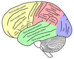
The inferior frontal gyrus is a gyrus of the frontal lobe (the yellow area of the human brain image to the right). It is labelled gyrus frontalis inferior, its Latin name. In the yellow area, its superior border is the inferior frontal sulcus (which divides it from the gyrus frontalis medius in the yellow area), its inferior border the lateral fissure (which divides it from the gyrus temporalis superior in the green area), and its posterior border is the inferior precentral sulcus (in the yellow area). Above it is the middle frontal gyrus (the gyrus frontalis medius), behind it the precentral gyrus (the gyrus praecentralis), both gyri in the yellow area of the image.[1]
The inferior frontal gyrus, like the middle frontal gyrus and the superior frontal gyrus, is more of a region than a true gyrus.
Structure
The inferior frontal gyrus can be subdivided into the following macroanatomical structures, shown in yellow in the top image, just below the label gyrus frontalis inferior:
- Pars opercularis (cortex posterior to the ascending ramus of the lateral fissure)
- Pars triangularis (cortex between the ascending ramus and the horizontal ramus of the lateral fissure)
- Pars orbitalis (cortex inferior and anterior to the horizontal ramus of the lateral fissure)
The inferior frontal gyrus includes the following cytoarchitectonic areas:
- Brodmann area 44
- Brodmann area 45
- Brodmann area 47
- cytoarchitectonic areas of the deep frontal operculum
The cytoarchitectonic areas very roughly correspond to the following macroanatomic structures: Brodmann area 44 to Pars opercularis, Brodmann area 45 to Pars triangularis, and Brodmann area 47 to Pars orbitalis. Brodmann area 44 corresponds to Broca's area (sometimes Broca's area is taken to encompass Brodmann's areas 44 and 45) — for the dominant hemisphere of the brain.
Language Processing
The left posterior inferior frontal gyrus (pIFG) is a part of the Articulatory Network I involved in motor syllable programs. The articulatory network itself contains three cortical areas; the posterior inferior frontal gyrus, the premotor cortex, and the anterior insula. These systems are interrelated but each has specific independent functions in speech comprehension and production. This system acts mostly when the vocal tract opens and closes during syllable production. Considered a "controller" of the motor aspect of speech production, the pIFG does not directly interact with the vocal tract; instead, it acts indirectly through the motor cortex. The posterior inferior frontal gyrus, connected to Brodmann Area 44, codes motor programs for this system while the auditory cortex (via the Spt) houses a series of sensory targets. Together, these areas function as a sensory-motor loop for syllable information coding.
In a study conducted comparing phonological and arithmetic processing and the involvement of different sections of the interior frontal gyrus and angular gyrus, cortical activation for phonology, subtraction, and multiplication tasks was compared. The predetermined language-calculation network was limited to the left inferior frontal gyrus, angular gyrus, superior parietal lobule, and the horizontal portion of the intraparietal sulcus. The results were significant to support that there was a pattern of left lateralization for each of these tasks all activating the Perisylvian fissure network, with some general localized areas for phonology and arithmetic. It was supported that phonology activated the pIFG and anterior angular gyrus, multiplication mainly implicated the anterior inferior frontal gyrus and the posterior angular gyrus. These systems are activated through similar neuronal processes but independently placed along the network.
Function
The left inferior frontal gyrus (IFG) is also extremely important for language comprehension and production due to the fact that most language processing takes place in the left hemisphere. Commonly known as "Broca's Area",[2] persons with damage in this region often have a type of non-fluent aphasia known as Broca's aphasia. Broca's area is located on the left hemisphere of the brain and encompasses Brodmann's area 44 and 45. Both overall have contributions to verbal fluency, but each has their own specific contribution. Area 44 is involved in the language production and phonological processing due to its connections with motor areas like the mouth and tongue. Area 45 is a part of the anterior inferior frontal gyrus and is involved in semantic processing. Characteristics of Broca's aphasia include agrammatic speech, relatively good language comprehension, poor repetition, and difficulty speaking. Persons with Broca's aphasia do not have deficits in language comprehension; however, they speak mostly in short utterances of a few words at a time, compromised mostly of nouns. Their speech is limited to short sentences, and producing sound is a very difficult task for those affected. The left IFG has also been suggested to play a role in inhibitory processes, including the tendency to inhibit learning from undesirable information. For example, TMS to the left IFG has been shown to release such inhibition, increasing the ability to learn from undesirable information.[3]
The right IFG, also considered Brodmann Area 44, has been implicated in go/no go[4] tasks. In such a task, the participant encounters a preliminary task (for instance repeatedly pressing a button), and then must halt this task when the actual go/no go signal is presented, ultimately measuring a level of impulse control through inhibition of a prepotent response. It seems that the same area is also implicated in risk aversion: a study found that higher risk aversion correlated with higher activity at IFG.[5] This might be explained as an inhibition signal to accept a risky option. Disruption of activity of this area with Transcranial Magnetic Stimulation or Direct Current Stimulation (tDCS) indeed leads to change in risk attitudes, as behaviorally demonstrated by choices over risky outcomes.[6][7]
References
- ↑ Nolte (2002), The Human Brain, ISBN 0-323-01320-1 photos on p526 & p.546
- ↑ The "dominant inferior frontal convolution" —Fauci, et.al, eds. (1998), Harrison's Principles of Internal Medicine, 14th Edition, Companion Handbook, ISBN 0-07-021530-8. p.1055
- ↑ Sharot, T., Kanai, R., Marston, D., Korn, C. W., Rees, G. & Dolan, R.J. (2012) Selectively Altering Belief Formation in the Human Brain. Proceeding of the National Academy of Sciences, 109 (42), 17058-17062.
- ↑ Aron AR, Robbins TW, Poldrack RA (2004). "Inhibition and the right inferior frontal cortex.". Trends Cogn Sci. 8 (4): 170–177. doi:10.1016/j.tics.2004.02.010. PMID 15050513.
- ↑ Christopoulos, GI.; Tobler, PN.; Bossaerts, P.; Dolan, RJ.; Schultz, W. (Oct 2009). "Neural correlates of value, risk, and risk aversion contributing to decision making under risk.". J Neurosci. 29 (40): 12574–83. doi:10.1523/JNEUROSCI.2614-09.2009. PMC 2794196
 . PMID 19812332.
 - ↑ Knoch D, Gianotti LR, Pascual-Leone A, Treyer V, Regard M, Hohmann M, Brugger P (2006). "Disruption of right prefrontal cortex by low-frequency repetitive transcranial magnetic stimulation induces risk-taking behavior.". J Neurosci. 26 (24): 6469–6472. doi:10.1523/JNEUROSCI.0804-06.2006. PMID 16775134.
- ↑ Fecteau S, Pascual-Leone A, Zald DH, Liguori P, Théoret H, Boggio PS, Fregni F (2007). "Activation of prefrontal cortex by transcranial direct current stimulation reduces appetite for risk during ambiguous decision making.". J Neurosci. 27 (23): 6212–6218. doi:10.1523/JNEUROSCI.0314-07.2007. PMID 17553993.
Additional images
 Inferior frontal gyrus(red).
 Lateral surface of left cerebral hemisphere, viewed from above.
 Coronal section through anterior cornua of lateral ventricles. Inferior frontal gyrus is shown in yellow.
 Drawing to illustrate the relations of the brain to the skull.
- Lateral view of a human brain, main gyri labeled.
 Human brain - left and right hemispheres - superior-lateral view: Inferior frontal gyrus is labelled 6
 Cerebrum. Lateral view.Deep dissection.
 Cerebrum. Lateral view.Deep dissection.
 Cerebrum. Lateral view.Deep dissection.
| Wikimedia Commons has media related to Inferior frontal gyrus. |