Hyaline cartilage
| Hyaline cartilage | |
|---|---|
 | |
| Identifiers | |
| Code | TH H2.00.03.5.00015 |
Hyaline cartilage is glass-like (hyaline) but translucent. It is found on many joint surfaces. It is pearl-grey in color with firm consistency and has a considerable amount of collagen. It contains no nerves or blood vessels, and its structure is relatively simple.
Structure
Hyaline cartilage is covered externally by a fibrous membrane, called the perichondrium, except at the articular ends of bones and also where it is found directly under the skin, i.e. ears and nose. This membrane contains vessels that provide the cartilage with nutrition.
Hyaline cartilage matrix is mostly made up of type II collagen and chondroitin sulfate, both of which are also found in elastic cartilage.
Hyaline cartilage exists on the ventral ends of ribs; in the larynx, trachea, and bronchi; and on the articulating surfaces of bones. It gives those structures a definite but pliable form. Presence of collagen fibres makes such structures and joints strong but less elastic and movable only to a minor extent. It is the most prevalent form of cartilage. It also forms the embryonic skeleton and the skeleton of elasmobranch fishes.
Histology
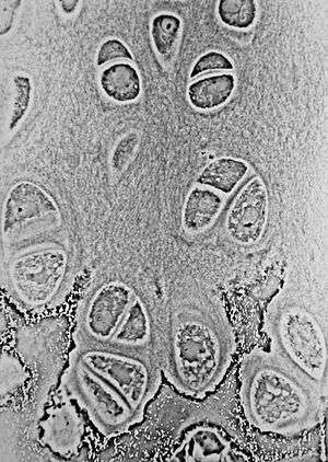
If a thin slice is examined under the microscope, it will be found to consist of cells (chondrocytes) of a rounded or bluntly angular form, lying in groups of two or more in a granular or almost homogeneous matrix. Chondrocytes are cartilage cells that produce the matrix. When arranged in groups of two or more, chondrocytes have generally straight outlines where they are in contact with each other, and in the rest of their circumference are rounded. They consist of clear translucent protoplasm in which fine interlacing filaments and minute granules are sometimes present; embedded in this are one or two round nuclei, having the usual intranuclear network.
The cells are contained in cavities in the matrix, called cartilage lacunae; these are actually artificial gaps formed by the shrinking of the cells during the staining and setting of the tissue for observation. The interterritorial space between the isogenous cell groups contains relatively more collagen fibers, causing it to maintain its shape while the actual cells shrink, creating the lacunae. This constitutes the so-called capsule of the space. Each lacuna is generally occupied by a single cell, but during the division of the cells it may contain two, four, or eight cells.
Articular cartilage
Articular cartilage is hyaline cartilage on the articular surfaces of bones.[1] As such, it lies inside the joint cavity of synovial joints, bathed in synovial fluid produced by the synovial membrane that lines the walls of the cavity.
Though it is often found in close contact with menisci and articular disks, articular cartilage is not considered a part of either of these structures, which are made entirely of fibrocartilage.
Additional images
 Articular cartilage of the elbow joint as seen in dissection
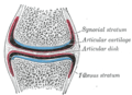 A synovial joint with bone, articular cartilage, and articular disc shown.
.jpg) Elbow joint. Deep dissection. Anterior view.
Elbow joint. Deep dissection. Anterior view.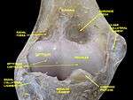 Elbow joint. Deep dissection. Anterior view..
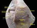 Elbow joint. Deep dissection. Posterior view.
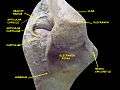 Elbow joint. Deep dissection. Posterior view.
See also
- Cartilage
- Hyaline
- Articular cartilage injuries
- Articular cartilage damage
- Articular cartilage repair
References
External links
| Wikimedia Commons has media related to hyaline cartilage.. |
- UIUC Histology Subject 331
- Histology image: 03301lba – Histology Learning System at Boston University