Horse hoof

A horse hoof is a structure surrounding the distal phalanx of the 3rd digit (digit III of the basic pentadactyl limb of vertebrates, evolved into a single weight-bearing digit in equids) of each of the four limbs of Equus species, which is covered by complex soft tissue and keratinised (cornified) structures. Since a single digit must bear the full proportion of the animal's weight that is borne by that limb, the hoof is of vital importance to the horse. The phrase "no hoof, no horse" underlines how much the health and the strength of the hoof is crucial for horse soundness.
Hooves in the natural state
Both wild and feral equid hooves have enormous strength and resilience, allowing any gait on any ground. A common example of the feral horse type is the Mustang. The Mustang is, in part, descended from the Iberian horses brought to the Americas by the Spanish, but most herds also have ancestry from other breeds. Therefore, the famous Mustang hoof strength is in part a result of natural selection and environment. Thus, it is proposed that other domestic breeds could develop similar hooves if raised under similar conditions.
The recent barefoot movement claims that such a strength can be almost completely restored to domesticated horses, when appropriate trimming and living conditions are applied, to such an extent that horseshoes are no longer necessary in almost any horse. If true, it would undermine the belief that "the horseshoe is a necessary evil."
The barefoot management system has not, however, gained a foothold among serious equine professionals, due to three factors: 1) increased strain placed on the hoof in sports, such as eventing and endurance riding, 2) the added weight of the rider and saddle, and 3) man-made surfaces, such as concrete, asphalt, and gravel, which can wear the walls down to the sensitive tissue over time.
Anatomy

The hoof is made up by an outer part, the hoof capsule (composed of various cornified specialised structures) and an inner, living part, containing soft tissues and bone. The cornified material of the hoof capsule is different in structure and properties in different parts. Dorsally, it covers, protects and supports P3 (also known as the coffin bone, pedal bone, PIII). Palmarly/plantarly, it covers and protects specialised soft tissues (tendons, ligaments, fibro-fatty and/or fibrocartilaginous tissues and cartilage). The upper, almost circular limit of the hoof capsule is the coronet (coronary band), having an angle to the ground of roughly similar magnitude in each pair of feet (i.e. fronts and backs). These angles may differ slightly from one horse to another, but not markedly. The walls originate from the coronet band. Walls are longer in the dorsal portion of the hoof (toe), intermediate in length in the lateral portion (quarter) and very short in palmar/plantar portion (heel). Heels are separated by an elastic, resilient structure named the 'frog'. In the palmar/plantar part of the foot, above the heels and the frog, there are two oval bulges named the 'bulbs'.
When viewed from the lower surface, the hoof wall's free margin encircles most of the hoof. The triangular frog occupies the center area. Lateral to the frog are two grooves, deeper in their posterior portion, named 'collateral grooves'. At the heels, the palmar/plantar portion of the walls bend inward sharply, following the external surface of collateral grooves to form the bars. The lower surface of the hoof, from the outer walls and the inner frog and bars, is covered by an exfoliating keratinised material, called the 'sole'.
Just below the coronet, the walls are covered for about an inch by a cornified, opaque 'periople' material. In the palmar/plantar part of the hoof, the periople is thicker and more rubbery over the heels, and it merges with frog material. Not all horses have the same amount of periople. Dry feet tend to lack this substance, which can be substituted with a hoof dressing.
Characters and functions of the external hoof structures
The walls
The walls are considered as a protective shield covering the sensitive internal hoof tissues (like the exoskeleton of arthropods), as a structure devoted to dissipating the energy of concussion, and as a surface to provide grip on different terrains. They are elastic and very tough, and vary in thickness from 6 to 12 mm. The walls are composed of three distinct layers: the pigmented layer, the water line and the white line.
The pigmented layer is generated by the coronet, and its color is just like that of the coronet skin from which it is derived. If the coronet skin has any dark patch, the walls show a parallel pigmented line, from the coronet to the ground, showing the wall's growth direction. This layer has predominately protective role, and is not as resistant to ground contact, where it can break and flake away.
The water line is built up by the coronet and by the wall's corium (the living tissue immediately beneath the walls). Its thickness increases proportionally to the distance from the coronet and, in the lower third of the walls, is thicker than the pigmented layer. It is very resistant to contact to the ground, and it serves mainly a support function.
The white line is the inner layer of the wall. It is softer and fibrous in structure and light in color; white in a freshly trimmed hoof, yellowish or gray after exposure to air and dirt. From the underside of the healthy hoof, it is seen as a thin line joining the sole and the walls. The white line grows out from the laminar connections. Any visible derangement of the white line indicates some important derangement of laminar connections that fix the walls to the underlying P3 bone. Since the white line is softer than both the walls and the sole, it wears fast where it appears on the surface; it appears as a subtle groove between the sole and the walls, often with some debris or sand inside.
The three layers of the wall merge in a single mass and they grow downwards together. If the wall does not wear naturally, from sufficient movement on abrasive terrains, then it will protrude from the solar surface. It then becomes prone to breakage, and the healthy hoof will self-trim, by breaking or chipping off.
When a horseshoe is applied, it is fixed to the wall. Nails are driven in, oblique to the walls. They enter the wall at the outside edge of the white line and they emerge at the wall's surface, about 15 to 20 mm from the base of the wall.
The wall is anatomically analogous to the human fingernail or toenail.
The frog
The frog is a V shaped structure that extends forwards across about two-thirds of the sole. Its thickness grows from the front to the back and, at the back, it merges with the heel periople. In its midline, it has a central groove (sulcus), that extends up between the bulbs.
It is dark gray-blackish in color and of a rubbery consistency, suggesting its role as shock absorber and grip tool on hard, smooth ground. The frog also acts like a pump to move the blood back to the heart, a great distance from the relatively thin leg to the main organ of the circulatory system.
In the stabled horse, the frog does not wear, but degrades, due to bacterial and fungal activity, to an irregular, soft, slashed surface. In the free-roaming horse, it hardens into a callous consistency with a near-smooth surface.
It is anatomically analogous to the human fingertip.
The sole
The sole has a whitish-yellowish, sometimes grayish color. It covers the whole space from the perimeter of the wall to the bars and the frog, on the underside of the hoof. Its deep layer has a compact, waxy character and it is called 'live sole'. Its surface is variable in character as a result of ground contact. If there is no contact, as in shod hooves or when the walls are too long or the movement poor, the lower surface of the sole has a crumbly consistency, and it is easily abraded by scratching it with a hoofpick. Conversely, it has a very hard consistency, with a smooth, bright surface, when there is a consistent, active contact with the ground. The front portion beneath the front of the pedal bone is called the 'sole callus'.
A stone bruise affects the sole of the horse's foot. It is often caused by a horse treading on a stone or sharp type of object, landings from high jumps and excessive exposure to snow. A major symptom is lameness.[1]
The bars
Bars are the inward folds of the wall, originating from the heels at an abrupt angle. The strong structure built up by the extremity of the heel and of the bar is named the 'heel buttress'. The sole between the heel walls and the bars is named the 'seat of corn', and it is a very important landmark used by natural hoof trimmers to evaluate the correct heel height. The bars have a three-layer structure, just like the walls (see above). When overgrown, they bend outwards and cover the lower surface of the sole.
Internal structures
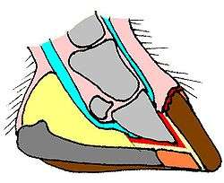
The third phalanx (coffin bone; pedal bone; P3;) is completely (or almost completely) covered by the hoof capsule. It has a crescent shape and a lower cup-like concavity. Its external surface mirrors the wall's shape. The corium, a dermo-epidermal, highly vascularized layer between the wall and the coffin bone, has a parallel, laminar shape, and is named the laminae. Laminar connection has a key role in the strength and the health of the hoof. Beneath the rear part of the sole, there is the digital cushion, which separates the frog and the bulb from underlying tendons, joints and bones, providing cushioning protection. In foals and yearlings, the digital cushion is composed of fibro-fatty, soft tissue. In the adult horse, it hardens into a fibro cartilagineous tissue when sufficient, consistent concussion stimulates the back of the hoof. Normal transformation of the digital cushion into fibrocartilagineous tissue is now considered a key goal, both for prevention of, and for rehabilitation of recovering cases of navicular syndrome . The flexor tendon lays deeper, just along the posterior surface of the small pastern bone (PII) and navicular bone, and it connects with posterior surface of P3; the navicular functions as a pulley.
The hoof mechanism
The horse hoof is not at all a rigid structure. It is elastic and flexible. Just squeezing the heels by hand will demonstrate that. When loaded, the hoof physiologically changes its shape. In part, this is a result of solar concavity, which has a variable depth, in the region of 1–1.5 cm. In part, it is a result of the arched shape of the lateral lower profile of the walls and sole, so that when an unloaded hoof touches a firm ground surface, there is only contact at toe and heels (active contact). A loaded hoof has a much greater area of ground contact (passive contact), covering the lower wall edge, most of the sole, bars and frog. Active contact areas can be seen as slightly protruding spots in the walls and in the callused sole.
The shape changes in a loaded hoof are complex. The plantar arch flattens, the solar concavity decreases in depth and heels spread. The hoof diameter increases to a 'dilated' configuration and P3 drops marginally into the hoof capsule. There is some recent evidence that a depression takes place in this phase, with blood pooling ('diastolic phase') mainly into the wall corium. When unloaded, the hoof restores its 'contracted' configuration, the pressure rises and the blood is squeezed out ('systolic phase'). There is a secondary pumping action, with the flexion of the foot, as it is raised.
The hoof mechanism ensures an effective blood circulation into the hoof, and it aids general circulation, too.
Time-related changes of the hoof
Hooves have to be considered as a plastic structure and their time-related, very complex changes can be considered in the short term (days/weeks), in the medium term (the horse's lifespan) and in the long term (the evolution of equids).
Hoof changes in the short term
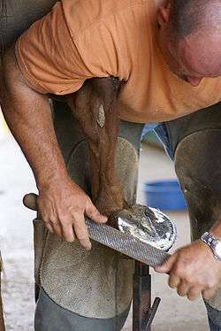
Just like the cornified layer of epidermis and of any mammalian nail, the hoof capsule is created only from epidermis, the outer living layer of the skin. From a microscopic point of view, epidermis is a multi-layered, specialised cornifying epithelium. It overlays the dermis, and it is separated from it by a basal lamina. It has no blood vessels and living cells acquire their oxygen and nutrients by fluid exchanges and molecular diffusion, from underlying dermis, flowing into microscopical spaces among individual cells. Products of metabolism are cleared by a reverse of this process. Epidermis growth take place by mitotic activity in its deepest layer, into the basal layer, with slow outward migration and maturation of cells. As these cells approach the surface, special proteins accumulate into their cytoplasm, then the cells die and 'dry', into microscopic, tightly-connected individual layers, composed mainly of keratin. The resulting 'dead' superficial layer serves a protective function, saving underlying living tissues from injury, from dehydration and from fungal and bacterial attack. The constant thickness of the cornified layer results most commonly from regular superficial exfoliation. When a specialised cornified structure has a particular toughness, as in nails and hair, little or no exfoliation occurs and the cornified structures must slowly migrate away from their original position.
Thus, the specialised cornified structures of the hoof are the wall, the sole, the frog and periople. The wall does not exfoliate at all; it is constantly growing downward (about 1 cm per month), and self-trims by wearing or chipping by ground contact, in wild and feral horses. Solar, frog and periople material grow outwards and exfoliate at the surface by ground contact and wearing. In the domesticated horse, movement and typical ground hardness are insufficient to allow self-trimming, so humans have to care for them, trimming the walls and the frog, and scraping off the dead sole.
Hoof changes in the medium term
Front and hind hooves are identical in the foal, but differ visibly in the adult horse. This is good evidence of medium-term plasticity of the whole hoof shape, as a result of variation in its use. Slow changes in hoof shape occur under any consistent change in the horse's movement pattern and under a wide variety of pathological conditions. They can be seen now as a clear example of a complex adaptive system, a frequent feature of living beings and structures.
Self-adapting capabilities of the hooves show their maximal effectiveness in wild equids (but domesticated horses show this too, to a lesser extent), as shown by the perfect soundness of feral horses, such as Mustangs, in a wide variety of environments.
Hoof changes in the long term
Equid hooves are the result of the 55-million-year evolution of the horse. Wild and domesticated Equus species share a very similar hoof shape and function. The present-day conformation of the hoof is a result of a progressive evolutionary loss of digits I, II, IV and V of the basal pentadactyl limb, with changes in bones, joints and hoof capsule. The resulting conformation allows a heavy, strong body to move with high speed on any ground, and most efficiently on open, hard, flat areas like prairies and deserts (i.e., 'cursorial specialisation').
Disorders
There are several disorders and injuries that can affect the equine hoof. Laminitis and navicular disease are two of the most serious. Thrush and white line disease, common bacterial infections, can become serious if left untreated. Quiltor, an infection of the lower leg that can travel under the hoof, is also sometimes seen, although most commonly in draft horses.
Quarter cracks are vertical splits in a hoof wall, most commonly seen on the inside of the front hooves or the outside of the hind hooves. They can result from poor shoeing and management practices, natural hoof conformation, or injuries to the leg and hoof.[2]
See also
External links
| Wikimedia Commons has media related to Horse hoof. |
- Horse Hoof Care Information Horse Hoof Care Net (CA)
- American Farrier's Association American Farrier's Association
- How a Horse Hoof Grows from eXtension
References
- ↑ "Stone Bruises Common in Thoroughbreds". Blood-Horse. Retrieved 19 July 2011.
- ↑ Reap, Stacey (December 26, 2008). "Resolving Quarter Cracks Takes More than Just a Stitch in Time". Chronicle of the Horse. Retrieved 2013-03-19.