History of anatomy
The history of anatomy extends from the earliest examinations of sacrificial victims to the sophisticated analyses of the body performed by modern scientists. It has been characterized, over time, by a continually developing understanding of the functions of organs and structures in the body.
Ancient anatomy
Egypt
The study of anatomy begins at least as early as 1600 BC, the date of the Edwin Smith Surgical Papyrus. This treatise shows that the heart, its vessels, liver, spleen, kidneys, hypothalamus, uterus and bladder were recognized, and that the blood vessels were known to emanate from the heart. Other vessels are described, some carrying air, some mucus, and two to the right ear are said to carry the "breath of life", while two to the left ear the "breath of death".The Ebers Papyrus (c. 1550 BC) features a treatise on the heart. It notes that the heart is the center of blood supply, and attached to it are vessels for every member of the body. The Egyptians seem to have known little about the function of the kidneys and made the heart the meeting point of a number of vessels which carried all the fluids of the body – blood, tears, urine and semen. However, they did not have a theory as to where saliva and sweat came from.[1]
Greek advances in anatomy
Nomenclature, methods and applications for the study of anatomy all date back to the Greeks.[2] The early scientist Alcmaeon began to construct a background for medical and anatomical science with the dissection of animals. He identified the optic nerves and the tubes later termed the Eustachius.[3] Others such as Acron (480 BC), Pausanias (480 BC), and Philistion of Locri made investigations into anatomy. One important figure during this time was Empedocles (480B.C.) who viewed the blood as the innate heat which he acquired from previous folklore. He also argued that the heart was the chief organ of both the vascular system and the pneuma (this could refer to either breath or soul; it was considered to be distributed by the blood vessels).[4]
Many medical texts by various authors are collected in the Hippocratic Corpus, none of which can definitely be ascribed to Hippocrates himself. The texts show an understanding of musculoskeletal structure, and the beginnings of understanding of the function of certain organs, such as the kidneys. The tricuspid valve of the heart and its function is documented in the treatise On the Heart.
In the 4th century BCE, Aristotle and several contemporaries produced a more empirically founded system, based on animal dissection. Through his work with animal dissections and evolutionary biology, Aristotle founded comparative anatomy. Around this time, Praxagoras is credited as the first to identify the difference between arteries and veins, and the relations between organs are described more accurately than in previous works.
The first recorded school of anatomy was in Alexandria from about 300 to the 2nd century BC.[5] Ptolemy I Soter was the first to allow for medical officials to cut open and examine dead bodies for the purposes of learning how human bodies operated. On some occasions King Ptolemy even took part in these dissections. Most of the early dissections were done on executed criminals. The first use of human cadavers for anatomical research occurred later in the 4th century BCE when Herophilos and Erasistratus gained permission to perform live dissections, or vivisection, on criminals in Alexandria under the auspices of the Ptolemaic dynasty. Herophilos in particular developed a body of anatomical knowledge much more informed by the actual structure of the human body than previous works had been. Herophilos was the first physician to dissect human bodies and is considered to be the founder of Anatomy. He reversed the longstanding notion made by Aristotle that the heart was the "seat of intelligence." He argued instead that this seat was the brain.[6] However, Herophilos was eventually accused by his contemporaries of dissecting live criminals. The number of victims is said to be around 600 prisoners.[7]
From ancient to medieval
Galen
The final major anatomist of ancient times was Galen, active in the 2nd century.[5] He compiled much of the knowledge obtained by previous writers, and furthered the inquiry into the function of organs by performing vivisection on animals. Due to a lack of readily available human specimens, discoveries through animal dissection were broadly applied to human anatomy as well. Galen served as chief physician to the gladiators in Pergamum (AD 158). Through his position with the gladiators, Galen was able to study all kinds of wounds without performing any actual human dissection. By default, Galen was able to view much of the abdominal cavity. His study on pigs and apes, however, gave him more detailed information about the organs and provided the basis for his medical tracts. Around 100 of these tracts survive and fill 22 volumes of modern text. His two great anatomical works are On anatomical procedure and On the uses of the parts of the body of man.[8] The information in these tracts became the foundation of authority for all medical writers and physicians for the next 1300 years until they were challenged by Vesalius and Harvey in the 16th century.[9][10]
It was through his experiments that Galen was able to overturn many long-held beliefs, such as the theory that the arteries contained air which carried it to all parts of the body from the heart and the lungs. This belief was based originally on the arteries of dead animals, which appeared to be empty. Galen was able to demonstrate that living arteries contain blood, but in his error, which became the established medical orthodoxy for centuries, was to assume that the blood goes back and forth from the heart in an ebb-and-flow motion.
Early modern anatomy

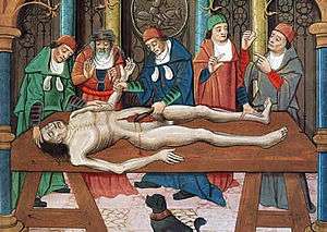
From the third century B.C.E until the twelfth century C.E. human anatomy was mainly learned through books and animal dissection.[11] While it was claimed by nineteenth century polemicists that human dissection became restricted after Boniface VIII passed a papal bull that forbade the dismemberment and boiling of corpses for funerary purposes and this is still repeated in some generalist works, this claim has been debunked as a myth by modern historians of science.[12] For many decades human dissection was thought unnecessary when all the knowledge about a human body could be read about from early authors such as Galen.[13] In The twelfth century as universities were being established in Italy, Emperor Frederick ii made it mandatory for students of medicine to take courses on human anatomy and surgery.[14] In the universities the lectern would sit elevated before the audience and instruct someone else in the dissection of the body, but in his early years Mondino de Luzzi performed the dissection himself making him one of the first and few to use a hands on approach to teaching human anatomy.[15]
Mondino de Luzzi "Mundinus" was born around 1276 and died in 1326,from 1314 to 1324 he presented many lectures on human anatomy at Bologna university.[16] Mondino de'Luzzi put together a book called "Anathomia" in 1316 that consisted of detailed dissections that he had performed, this book was used as a text book in universities for 250 years.[17] "Mundinus" carried out the first systematic human dissections since Herophilus of Chalcedon and Erasistratus of Ceos 1500 years earlier.[18][19] The first major development in anatomy in Christian Europe since the fall of Rome occurred at Bologna, where anatomists dissected cadavers and contributed to the accurate description of organs and the identification of their functions. Following de Liuzzi's early studies, fifteenth century anatomists included Alessandro Achillini and Antonio Benivieni [18][20] Pathological anatomy[21]
Leonardo da Vinci
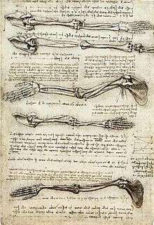
Leonardo da Vinci (1452–1519) was trained in anatomy by Andrea del Verrocchio. In 1489 Leonardo began a series of anatomical drawings depicting the ideal human form. This work was carried out intermittently for over 2 decades. During this time he made use of his anatomical knowledge in his artwork, making many sketches of skeletal structures, muscles and organs of humans and other vertebrates that he dissected.[22][23] Initially adopting an Aristotlean understanding of anatomy, he later studied Galen and adopted a more empirical approach, eventually abandoning Galen altogether and relying entirely on his own direct observation.[24] His surviving 750 drawings represent groundbreaking studies in anatomy. Leonardo dissected around thirty human specimens until he was forced to stop under order of Pope Leo X.
As an artist-anatomist, Leonardo made many important discoveries, and had intended to publish a comprehensive treatise on human anatomy.[24] For instance, he produced the first accurate depiction of the human spine, while his notes documenting his dissection of the Florentine centenarian contain the earliest known description of cirrhosis of the liver and arteriosclerosis.[24][25] He was the first to develop drawing techniques in anatomy to convey information using cross-sections and multiple angles, although centuries would pass before anatomical drawings became accepted as crucial for learning anatomy.[26] None of Leonardo's Notebooks were published during his lifetime, many being lost after his death, with the result that his anatomical discoveries remained unknown until they were later found and published centuries after his death.[27]
Vesalius
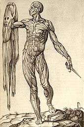
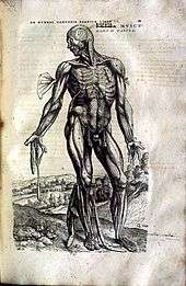
The Galenic doctrine in Europe was first seriously challenged in the 16th century. Thanks to the printing press, all over Europe a collective effort proceeded to circulate the works of Galen and later publish criticisms on their works. Andreas Vesalius, born and educated in Belgium, contributed the most to human anatomy. Vesalius was the first to publish a treatise, De humani corporis fabrica, that challenged Galen "drawing for drawing." These drawings were a detailed series of explanations and vivid drawings of the anatomical parts of human bodies. Vesalius traveled all the way from Leuven[28] to Padua for permission to dissect victims from the gallows without fear of persecution. His superbly executed drawings are triumphant descriptions of the differences between dogs and humans, but it took a century for Galen's influence to fade. His work led to anatomy marked a new era in the study of anatomy and its relation to medicine. Under Vesalius, anatomy became an actual discipline. “His skill in and attention to dissection featured prominently in his publications as well as his demonstrations, in his research as well as his teaching." [29] In 1540, Vesalius gave a public demonstration of the inaccuracies of Galen's anatomical theories, which are still the orthodoxy of the medical profession. Vesalius now has on display, for comparison purposes, the skeletons of a human being alongside that of an ape of which he was able to show, that in many cases, Galen's observations were indeed correct for the ape, but bear little relation to man. Clearly what was needed was a new account of human anatomy. While the lecturer explained human anatomy, as revealed by Galen more than 1000 years earlier, an assistant pointed to the equivalent details on a dissected corpse. At times, the assistant was unable to find the organ as described, but invariably the corpse rather than Galen was held to be in error. Vesalius then decided that he will dissect corpses himself and trust to the evidence of what he found. His approach was highly controversial, but his evident skill led to his appointment as professor of surgery and anatomy at the University of Padua.
A succession of researchers proceeded to refine the body of anatomical knowledge, giving their names to a number of anatomical structures along the way. The 16th and 17th centuries also witnessed significant advances in the understanding of the circulatory system, as the purpose of valves in veins was identified, the left-to-right ventricle flow of blood through the circulatory system was described, and the hepatic veins were identified as a separate portion of the circulatory system. The lymphatic system was also identified as a separate system at this time.
17th and 18th centuries
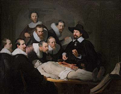
The study of anatomy flourished in the 17th and 18th centuries. The advent of the printing press facilitated the exchange of ideas. Because the study of anatomy concerned observation and drawings, the popularity of the anatomist was equal to the quality of his drawing talents, and one need not be an expert in Latin to take part. Many famous artists studied anatomy, attended dissections, and published drawings for money, from Michelangelo to Rembrandt. For the first time, prominent universities could teach something about anatomy through drawings, rather than relying on knowledge of Latin. Contrary to popular belief, the Church neither objected to nor obstructed anatomical research.[30]
Only certified anatomists were allowed to perform dissections, and sometimes then only yearly. These dissections were sponsored by the city councilors and often charged an admission fee, rather like a circus act for scholars. Many European cities, such as Amsterdam, London, Copenhagen, Padua, and Paris, all had Royal anatomists (or some such office) tied to local government. Indeed, Nicolaes Tulp was Mayor of Amsterdam for three terms. Though it was a risky business to perform dissections, and unpredictable depending on the availability of fresh bodies, attending dissections was legal.
To cope with shortages of cadavers and the rise in medical students during the 17th and 18th centuries, body-snatching and even anatomy murder were practiced to obtain cadavers.[31] 'Body snatching' was the act of sneaking into a graveyard, digging up a corpse and using it for study. Men known as 'resurrectionists' emerged as outside parties, who would steal corpses for a living and sell the bodies to anatomy schools. The leading London anatomist John Hunter paid for a regular supply of corpses for his anatomy school.[32] The British Parliament passed the Anatomy Act 1832, which finally provided for an adequate and legitimate supply of corpses by allowing legal dissection of executed murderers. The view of anatomist at the time, however, became similar to that of an executioner. Having one's body dissected was seen as a punishment worse than death, "if you stole a pig, you were hung. If you killed a man, you were hung and then dissected." Demand grew so great that some anatomist resorted to dissecting their own family members (William Harvey dissected his own father and sister) as well as robbing bodies from their graves.[33]
Many Europeans interested in the study of anatomy traveled to Italy, then the centre of anatomy. Only in Italy could certain important research methods be used, such as dissections on women. Realdo Colombo (also known as Realdus Columbus) and Gabriele Falloppio were pupils of Vesalius. Columbus, as Vesalius's immediate successor in Padua, and afterwards professor at Rome, distinguished himself by describing the shape and cavities of the heart, the structure of the pulmonary artery and aorta and their valves, and tracing the course of the blood from the right to the left side of the heart.[34]
Anatomical theatres

Anatomical theatres became a popular form for anatomical teaching in the early 16th century. The University of Padua was the first and most widely known theatre, founded in 1594. As a result, Italy became the center for human dissection. People came from all over to watch as professors taught lectures on the human physiology and anatomy, as anyone was welcome to witness the spectacle. Participants “were fascinated by corporeal display, by the body undergoing dissection.”.[35] Most professors did not do the dissections themselves. Instead they sat in seats above the bodies while hired hands did the cutting. Students and observers would be placed around the table in a circular, stadium like arena and listen as professors explained the various anatomical parts. The 19th century eventually saw a move from anatomical theatres to classrooms, reducing "the number of people who could benefit from each cadaver." [5]
19th century anatomy
During the 19th century, anatomical research was extended with histology and developmental biology of both humans and animals. Women, who were not allowed to attend medical school, could attend the anatomy theatres. From 1822 the Royal College of Surgeons forced unregulated schools to close.[36] Medical museums provided examples in comparative anatomy, and were often used in teaching.[37]
Modern anatomy

Anatomical research in the past hundred years has taken advantage of technological developments and growing understanding of sciences such as evolutionary and molecular biology to create a thorough understanding of the body's organs and structures. Disciplines such as endocrinology have explained the purpose of glands that anatomists previously could not explain; medical devices such as MRI machines and CAT scanners have enabled researchers to study organs, living or dead, in unprecedented detail. Progress today in anatomy is centered in the development, evolution, and function of anatomical features, as the macroscopic aspects of human anatomy have largely been catalogued. Non-human anatomy is particularly active as researchers use techniques ranging from finite element analysis to molecular biology.
To save time, some medical schools such as Birmingham, England have adopted prosection, where a demonstrator dissects and explains to an audience, in place of dissection by students. This enables students to observe more than one body. Improvements in colour images and photography means that an anatomy text is no longer an aid to dissection but rather a central material to learn from. Plastic models are regularly used in anatomy teaching, offering a good substitute to the real thing. Use of living models for anatomy demonstration is once again becoming popular within teaching of anatomy. Surface landmarks that can be palpated on another individual provide practice for future clinical situations. It is possible to do this on oneself; in the Integrated Biology course at the University of Berkeley, students are encouraged to "introspect"[38] on themselves and link what they are being taught to their own body.[36]
Donations of bodies have declined with public confidence in the medical profession.[39] In Britain, the Human Tissue Act 2004 has tightened up the availability of resources to anatomy departments. The outbreaks of Bovine Spongiform Encephalitis (BSE) in the late 80s and early 90s further restricted the handling of brain tissue.[36][40]
The controversy with Gunther von Hagens and public displays of dissections, preserved by plastination, may divide opinions on what is ethical or legal.[41]
References
- ↑ Porter, Roy (1999-10-17). The Greatest Benefit to Mankind: A Medical History of Humanity (The Norton History of Science). W. W. Norton. pp. 49–50. ISBN 9780393319804. Retrieved 17 November 2013.
- ↑ Singer, Charles (1957). A Short History of Anatomy & Physiology from Greeks to Harvey. NEw York: Dover Publications Inc. p. 5.
- ↑ Singer, Charles (1957). A Short History of Anatomy & Physiology from Greeks to Harvey. NEw York: Dover Publications Inc. p. 7.
- ↑ Singer, Charles (1957). A Short History of Anatomy & Physiology from Greeks to Harvey. NEw York: Dover Publications Inc. p. 10.
- 1 2 3 Siddiquey, Ak Shamsuddin Husain (2009). "History of Anatomy". Bangladesh Journal of Anatomy. 7 (1).
- ↑ Singer, Charles (1957). A Short History of Anatomy & Physiology from Greeks to Harvey. NEw York: Dover Publications Inc. p. 29.
- ↑ Roach, Mary (2003). Stiff: The curious Lives of Human Cadavers. New York: W.W. Norton. p. 41.
- ↑ Singer, Charles (1957). A Short History of Anatomy & Physiology from Greeks to Harvey. NEw York: Dover Publications Inc. p. 47.
- ↑ Boas, Marie (1970). The Scientific Renaissance 1450-1630. Fontana. pp. 120, 248.
Vesalius, finding Galen full of errors, was quite certain that he had been able to eradicate them.
- ↑ Boas, Marie (1970). The Scientific Renaissance 1450-1630. Fontana. p. 262.
Like any sixteenth-century anatomist too he [Harvey] began with what Gelen had taught, and managed to interpret Galen's words to win support for his new doctrine.
- ↑ Siraisi, Nancey G. (1990). Medieval and Early Renaissance Medicine. Chicago and London: University of Chicago Press. p. 84. ISBN 0-226-76129-0.
- ↑ Park, Katherine (November 2010). "Myth 5 - That the Medieval Church Prohibited Dissection". In Numbers, Ronald L. Galileo Goes to Jail and Other Myths about Science and Religion. Harvard University Press. pp. 43–49. ISBN 9780674057418.
- ↑ Nutton, Vivian (2004). Ancient Medicine. London and New York: Routledge Taylor & Francis Group. p. 138. ISBN 978-0-415-36848-3.
- ↑ Crombie, A.C. (1967). Medieval and Early Modern Science (volume 1 ed.). Cambridge, Massachusetts: Harvard University Press. p. 180 and 181.
- ↑ Persaud, Tubbs, Loukas, T.V.N, Shane R. Marios (2014). A History of Human Anatomy. Springfield, Illinois: Charles C. Thomas Publisher, LTD. p. 55. ISBN 978-0-398-08105-8.
- ↑ Gordon, Benjamin Lee (1959). Medieval and Renaissance Medicine. New York: Philosophical Library, Inc. pp. 422–426.
- ↑ Persaud, Loukas, Tubbs, T.V.N. Marios, Shane R. (2014). A History of Human Anatomy (Second ed.). Springfield, Illinois, U.S.A: Charles C. Thomas, Publisher, LTD. pp. 56, 55–59. ISBN 978-0-398-08105-8. Retrieved 2015-11-28.
- 1 2 Zimmerman, Leo M.; Veith, Ilza (1993-08-01). Great Ideas in the History of Surgery. Norman Publishing. ISBN 9780930405533. Retrieved 7 December 2012.
- ↑ Crombie, Alistair Cameron (1959). The History of Science From Augustine to Galileo. Courier Dover Publications. ISBN 9780486288505. Retrieved 19 December 2012.
- ↑ Benivieni, Antonio; Polybus; Guinterius, Joannes (1529). De abditis nonnullis ac mirandis morborum & sanationum causis. apud Andream Cratandrum. Retrieved 7 December 2012.
- ↑ Thorndike, Lynn (1958). A History of Magic and Experimental Science: Fourteenth and fifteenth centuries. Columbia University Press. ISBN 9780231087971. Retrieved 7 December 2012.
- ↑ Boas, Marie (1970). The Scientific Renaissance 1450–1630. Fontana. pp. 120–143. (First published by Collins, 1962.)
- ↑ Mason, Stephen F. (1962). A History of the Sciences. New York: Collier. p. 550.
- 1 2 3 O'Malley, Charles D. (1983). Leonardo on the Human Body. New York: Dover.
- ↑ "Leonardo the Man , His machines". Lairweb. Retrieved 2 November 2014.
- ↑ "Leonardo Da Vinci first Anatomist". Life in The Fast Lane. Retrieved 2 November 2014.
- ↑ "Leonardo Da Vinci's Notebook Project". Irvine Valley College. Retrieved 2 November 2014.
- ↑ "Katholieke Universiteit Leuven". Kuleuven.ac.be. Retrieved 2012-06-13.
- ↑ Klestinec, Cynthia (2004). "A History of Anatomy Theaters in Sixteenth-Century Padua". Journal of the History of Medicine and Allied Sciences. 59 (3): 375–412. doi:10.1093/jhmas/59.3.375.
- ↑ Howse, Christopher (10 June 2009). "The myth of the anatomy lesson". The Daily Telegraph. London. Retrieved 4 May 2010.
- ↑ Rosner, Lisa. 2010. The Anatomy Murders. Being the True and Spectacular History of Edinburgh's Notorious Burke and Hare and of the Man of Science Who Abetted Them in the Commission of Their Most Heinous Crimes. University of Pennsylvania Press
- ↑ Moore, Wendy (2006). The Knife Man: Blood, Body-Snatching and the Birth of Modern Surgery. Bantam. pp. 87–95 and passim. ISBN 0-553-81618-7.
- ↑ Roach, Mary (2003). Stiff: The curious Lives of Human Cadavers. New York: W.W. Norton. pp. 37–57.
- ↑ Boas, Marie (1970). The Scientific Renaissance 1450-1630. Fontana. pp. 254–256.
- ↑ Klestinec, Cynthia (2004). "A History of Anatomy Theaters in Sixteenth-Century Padua". Journal of the History of Medicine and Allied Sciences. 59 (3): 375–412. doi:10.1093/jhmas/59.3.375.
- 1 2 3 McLachlan J.; Patten D. (2006). "Anatomy teaching: ghosts of the past, present and future". Medical Education. 40 (3): 243–53. doi:10.1111/j.1365-2929.2006.02401.x.
- ↑ Reinarz J (2005). "The age of museum medicine: The rise and fall of the medical museum at Birmingham's School of Medicine". Social History of Medicine. 18 (3): 419–37. doi:10.1093/shm/hki050.
- ↑ Diamond M. 2005. Integrative Biology 131 - Lecture 01: Organization of Body. Berkeley, University of California.
- ↑ British Broadcasting Corporation (BBC) News. 2001. Organ scandal background. http://news.bbc.co.uk/1/hi/health/1136723.stm Accessed 22 April 2008.
- ↑ Demiryurek D. Bayramoglu; Ustacelebi S. (2002). "Infective agents in fixed human cadavers: a brief review and suggested guidelines". Anatomical Record. 269 (1): 194–7.
- ↑ British Broadcasting Corporation (BBC) News. 2002 Controversial autopsy goes ahead. http://news.bbc.co.uk/1/hi/health/2493291.stm Accessed 22 April 2008.
Bibliography
| Wikimedia Commons has media related to History of anatomy. |
- Knoeff, Rina (2012). Dutch Anatomy and Clinical Medicine in 17th-Century Europe. Leibniz Institute of European History.
- Mazzio, C. (1997). The Body in Parts: Discourses and Anatomies in Early Modern Europe. Routledge. ISBN 0-415-91694-1.
- Porter, R. (1997). The Greatest Benefit to Mankind: A Medical History of Humanity from Antiquity to the Present. Harper Collins. ISBN 0-00-215173-1.
- Sawday, J. (1996). The Body Emblazoned: Dissection and the Human Body in Renaissance Culture. Routledge. ISBN 0-415-15719-6.
External links
- Historical Anatomies on the Web. National Library of Medicine. Selected images from notable anatomical atlases.
- Anatomia 1522-1867: Anatomical Plates from the Thomas Fisher Rare Book Library
- Human Anatomy & Physiology Society A society to promote communication among teachers of human anatomy and physiology in colleges, universities, and related institutions.
| Histories of basic sciences |  | |
|---|---|---|
| Histories of medical specialties | ||
| Medicine in ancient societies | ||
| History of methods in medicine | ||
| Disasters and plagues | ||
| ||