Hiatus hernia
| Hiatus hernia | |
|---|---|
 | |
| Classification and external resources | |
| Specialty | Gastroenterology, general surgery |
| ICD-10 | K44, Q40.1 |
| ICD-9-CM | 553.3, 750.6 |
| OMIM | 142400 |
| DiseasesDB | 29116 |
| MedlinePlus | 001137 |
| eMedicine | med/1012 radio/337 |
| MeSH | D006551 |
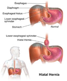
A hiatus hernia or hiatal hernia is the protrusion (or herniation) of the upper part of the stomach into the chest cavity through the esophageal hiatus because of a tear or weakness in the diaphragm.
The most common cause is obesity. The diagnosis is often by endoscopy or medical imaging.[1]
A hiatus hernia may be treated with lifestyle changes such as raising the head of the bed, weight loss, and adjusting eating habits. Medications such as H2 blockers or proton pump inhibitors may help. If the symptoms do not improve with medications the surgery known as laparoscopic fundoplication may be an option.[1]
Signs and symptoms

Hiatal hernia has often been called the "great mimic" because its symptoms can resemble many disorders. For example, a person with this problem can experience dull pains in the chest, shortness of breath (caused by the hernia's effect on the diaphragm), heart palpitations (due to irritation of the vagus nerve), and swallowed food "balling up" and causing discomfort in lower esophagus until it passes on to stomach. Hiatus hernias often result in heartburn but may also cause chest pain or pain with eating.[1]
In most cases however, a hiatal hernia does not cause any symptoms. The pain and discomfort that a patient experiences is due to the reflux of gastric acid, air, or bile. While there are several causes of acid reflux, it does happen more frequently in the presence of hiatal hernia.
In newborns, the presence of Bochdalek hernia can be recognised[2] from symptoms such as difficulty breathing[3] fast respiration, increased heart rate.[4]
Risk factors
The following are risk factors that can result in a hiatus hernia.
- Increased pressure within the abdomen caused by:
- Heavy lifting or bending over
- Frequent or hard coughing
- Hard sneezing
- Violent vomiting
- Straining
- Stress
Diagnosis
The diagnosis of a hiatus hernia is typically made through an upper GI series, endoscopy or high resolution manometry.
-
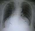
A large hiatus hernia on X-ray marked by open arrows in contrast to the heart borders marked by closed arrows
-

Upper GI endoscopy depicting hiatus hernia
-

A hiatus hernia as seen on CT
-

A large hiatus hernia as seen on CT imaging
-

A large hiatus hernia as seen on CT imaging
Classification

Four types of esophageal hiatal hernia are identified:[5]
Type I: A type I hernia is also known as a sliding hiatal hernia. There is a widening of the muscular hiatal tunnel and circumferential laxity of the phrenoesophageal membrane, allowing a portion of the gastric cardia to herniate upward into the posterior mediastinum. The clinical significance of type I hernias is in their association with reflux disease. Sliding hernias are the most common type, and account 95% of all hiatal hernias.[6] (C)
Type II: A type II hernia results from a localized defect in the phrenoesophageal membrane while the gastroesophageal junction remains fixed to the pre aortic fascia and the median arcuate ligament. The gastric fundus then serves as the leading point of herniation. Although type II hernias are associated with reflux disease, their primary clinical significance lies in the potential for mechanical complications. (D)
Type III: Type III hernias have elements of both types I and II hernias. With progressive enlargement of the hernia through the hiatus, the phrenoesophageal membrane stretches, displacing the gastroesophageal junction above the diaphragm, thereby adding a sliding element to the type II hernia.
Type IV: Type IV hiatus hernia is associated with a large defect in the phrenoesophageal membrane, allowing other organs, such as colon, spleen, pancreas and small intestine to enter the hernia sac.
The end stage of type I and type II hernias occurs when the whole stomach migrates up into the chest by rotating 180° around its longitudinal axis, with the cardia and pylorus as fixed points. In this situation the abnormality is usually referred to as an intrathoracic stomach.
Treatment
In the great majority of cases, sufferers experience no life-altering discomfort, and no treatment is required. If there is pain or discomfort, 3 or 4 sips of room temperature water will usually relieve the pain. Symptomatic patients should elevate the head of their beds and avoid lying down directly after meals. If the condition has been brought on by stress, stress reduction techniques may be prescribed, or if overweight, weight loss may be indicated. Antisecretory drugs like proton pump inhibitors and H2 receptor blockers can be used to reduce acid secretion. Medications that reduce the lower esophageal sphincter (LES) pressure should be avoided.
However in some unusual instances, as when the hiatal hernia is unusually large, or is of the paraesophageal type, it may cause esophageal stricture or severe discomfort. About 5% of hiatus hernias are paraesophageal. If symptoms from such a hernia are severe for example if chronic acid reflux threatens to severely injure the esophagus or is causing Barrett's esophagus, surgery is sometimes recommended. However surgery has its own risks including death and disability, so that even for large or paraesophageal hernias, watchful waiting may on balance be safer and cause fewer problems than surgery.[7] Complications from surgical procedures to correct a hiatus hernia may include gas bloat syndrome, dysphagia (trouble swallowing), dumping syndrome, excessive scarring, and rarely, achalasia.[8] Surgical procedures sometimes fails over time, requiring a second surgery to make repairs.
One surgical procedure used is called Nissen fundoplication. In fundoplication, the gastric fundus (upper part) of the stomach is wrapped, or plicated, around the inferior part of the esophagus, preventing herniation of the stomach through the hiatus in the diaphragm and the reflux of gastric acid. The procedure is now commonly performed laparoscopically. With proper patient selection, laparoscopic fundoplication recent studies have indicated relatively low complication rates, quick recovery, and relatively good long term results.[9][10][11][12][13]
Epidemiology
Incidence of hiatal hernias increases with age; approximately 60% of individuals aged 50 or older have a hiatal hernia.[14] Of these, 9% are symptomatic, depending on the competence of the lower esophageal sphincter (LES). 95% of these are "sliding" hiatus hernias, in which the LES protrudes above the diaphragm along with the stomach, and only 5% are the "rolling" type (paraesophageal), in which the LES remains stationary but the stomach protrudes above the diaphragm. People of all ages can get this condition, but it is more common in older people.
According to Dr. Denis Burkitt, "Hiatus hernia has its maximum prevalence in economically developed communities in North America and Western Europe ... In contrast the disease is rare in situations typified by rural African communities."[15] Burkitt attributes the disease to insufficient dietary fiber and the use of the unnatural sitting position for defecation. Both factors create the need for straining at stool, increasing intraabdominal pressure and pushing the stomach through the esophageal hiatus in the diaphragm.[16]
References
- 1 2 3 Roman, S; Kahrilas, PJ (23 October 2014). "The diagnosis and management of hiatus hernia.". BMJ (Clinical research ed.). 349: g6154. doi:10.1136/bmj.g6154. PMID 25341679.
- ↑ Chang SW, Lee HC, Yeung CY, Chan WT, Hsu CH, Kao HA, Hung HY, Chang JH, Sheu JC, Wang NL (2010). "A twenty-year review of early and late-presenting congenital Bochdalek diaphragmatic hernia: are they different clinical spectra?". Pediatr Neonatol. 51 (1): 26–30. doi:10.1016/S1875-9572(10)60006-X. PMID 20225535.
- ↑ doi: :10.1016/j.athoracsur.2013
- ↑ Alam, A; Chander, B.N. (July 2005). "Adult bochdalek hernia". MJAFI. 61 (3): 284–6. doi:10.1016/S0377-1237(05)80177-7.
- ↑ Kahrilas, Peter J.; Kim, Hyon C.; Pandolfino, John E. "Approaches to the diagnosis and grading of hiatal hernia". Best Practice & Research Clinical Gastroenterology. 22 (4): 601–616. doi:10.1016/j.bpg.2007.12.007. PMC 2548324
 . PMID 18656819.
 - ↑ Dennis Kasper, Anthony Fauci, Stephen Hauser, Dan Longo, J. Larry Jameson, Joseph Loscalzo. Harrison's Principles of Internal Medicine, 19e (19 ed.). McGraw-Hill Global Education. p. 1902. ISBN 978-0071802154.
- ↑ Paraesophageal Hernias: Operation or Observation? Ann Surg. 2002 Oct; 236(4): 492–501 PMCID: PMC1422604
- ↑ Paraesophageal Hernias: Operation or Observation? Ann Surg. 2002 Oct; 236(4): 492–501. PMCID: PMC1422604
- ↑ Migaczewski M, Pędziwiatr M, Matłok M, Budzyński A (2013). "Laparoscopic Nissen fundoplication in the treatment of Barrett's esophagus — 10 years of experience". Wideochir Inne Tech Maloinwazyjne. 8 (2): 139–45. doi:10.5114/wiitm.2011.32941. PMC 3699774. PMID 23837098.
- ↑ Witteman BP, Strijkers R, de Vries E, Toemen L, Conchillo JM, Hameeteman W, Dagnelie PC, Koek GH, Bouvy ND (2012). "Transoral incisionless fundoplication for treatment of gastroesophageal reflux disease in clinical practice". Surg Endosc. 26 (11): 3307–15. doi:10.1007/s00464-012-2324-2. PMC 3472060. PMID 22648098.
- ↑ Ozmen V, Oran ES, Gorgun E, Asoglu O, Igci A, Kecer M, Dizdaroglu F (2006). "Histologic and clinical outcome after laparoscopic Nissen fundoplication for gastroesophageal reflux disease and Barrett's esophagus". Surg Endosc. 20 (2): 226–9. doi:10.1007/s00464-005-0434-9. PMID 16362470.
- ↑ Abbas AE, Deschamps C, Cassivi SD, Allen MS, Nichols FC, Miller DL, Pairolero PC (2004). "Barrett's esophagus: the role of laparoscopic fundoplication". Ann. Thorac. Surg. 77 (2): 393–6. doi:10.1016/S0003-4975(03)01352-3. PMID 14759403.
- ↑ "Journal Index PDF (fee for article)" (PDF). Lange Current Medical Diagnosis & Treatment 2006.
- ↑ Goyal Raj K, "Chapter 286. Diseases of the Esophagus". Harrison's Principles of Internal Medicine, 17e.
- ↑ Burkitt DP (1981). "Hiatus hernia: is it preventable?" (PDF). Am. J. Clin. Nutr. 34 (3): 428–31. PMID 6259926.
- ↑ Sontag S (1999). "Defining GERD". Yale J Biol Med. 72 (2-3): 69–80. PMC 2579007. PMID 10780568.
External links
| Wikimedia Commons has media related to Hiatal hernia. |