Dirofilaria immitis
| Dirofilaria immitis | |
|---|---|
 | |
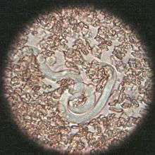
| A German Shepherd dog heart infested with Dirofilaria immitis | |
| Scientific classification | |
| Kingdom: | Animalia |
| Phylum: | Nematoda |
| Class: | Secernentea |
| Order: | Spirurida |
| Family: | Onchocercidae |
| Genus: | Dirofilaria |
| Species: | D. immitis |
| Binomial name | |
| Dirofilaria immitis (Leidy, 1856) | |
Dirofilaria immitis, the heartworm or dog heartworm, is a parasitic roundworm that is spread from host to host through the bites of mosquitoes. The heartworm is a type of filarial worm, a small thread-like worm, that causes filariasis. The definitive host is the dog, but it can also infect cats, wolves, coyotes, foxes, and other animals, such as ferrets, sea lions and even, under very rare circumstances, humans.[1] The parasite is commonly called "heartworm"; however, adults often reside in the pulmonary arterial system (lung arteries), as well as the heart, and a major effect on the health for the animal is a manifestation of damage to the lung vessels and tissues.[2] Occasionally, adult heartworms migrate to the right heart and even the great veins in heavy infections. Heartworm infection may result in serious disease for the host, with death typically as the result of congestive heart failure.
Distribution
Although at one time confined to the southern United States, heartworm has now spread to nearly all locations where its vector, the mosquito, is found. Transmission of the parasite occurs in all of the United States (cases have even been reported in Alaska), and the warmer regions of Canada. The highest infection rates are found within 150 miles of the coast from Texas to New Jersey, and along the Mississippi River and its major tributaries.[3] It has also been found in South America,[4] southern Europe,[5][6] Southeast Asia,[7] the Middle East,[8] Australia, Korea, and Japan.[3][9]
Course of infection

Heartworms go throughout several life stages before they become adults infecting the pulmonary artery of the host animal. The worms require the mosquito as an intermediate stage to complete their lifecycles. The rate of development in the mosquito is temperature-dependent, requiring about two weeks of temperature at or above 27°C (80°F). Below a threshold temperature of 14°C (57°F), development cannot occur, and the cycle is halted.[10] As a result, transmission is limited to warm weather, and duration of the transmission season varies geographically. The period between the initial infection when the dog is bitten by a mosquito and the maturation of the worms into adults living in the heart takes six to seven months in dogs and is known as the "prepatent period".
After infection, the third-stage larval heartworms (L3) deposited by the mosquito grow for a week or two and molt to the fourth larval stage (L4) under the skin at the site of the mosquito bite. Then, they migrate to the muscles of the chest and abdomen, and 45 to 60 days after infection, molt to the fifth stage (L5, immature adult). Between 75 and 120 days after infection, these immature heartworms then enter the bloodstream and are carried through the heart to reside in the pulmonary artery. Over the next three to four months, they increase greatly in size. The female adult worm is about 30 cm in length, and the male is about 23 cm, with a coiled tail.[11] By seven months after infection, the adult worms have mated and the females begin giving birth to live young, called microfilariae.
The microfilariae circulate in the bloodstream for as long as two years, waiting for the next stage in their lifecycles in the gut of a bloodsucking mosquito. When ingested by a mosquito, the microfilariae undergo a series of molts to the infective third larval stage, and then migrate to the salivary glands of the mosquito, where they wait to infect another host. The incubation period required to reach the stage where the microfilariae become transmittable to another host can be as little as two weeks or as long as six weeks, depending on the warmth of the climate, and the larval lifecycle ceases entirely if the ambient temperature drops below 14°C (57°F).
Hosts
Hosts of Dirofilaria immitis include:[1]
Clinical signs of infection
Dogs show no indication of heartworm infection during the six-month prepatent period prior to the worms' maturation, and current diagnostic tests for the presence of microfilariae or antigens cannot detect prepatent infections. Rarely, migrating heartworm larvae get "lost" and end up in unusual sites, such as the eye, brain, or an artery in the leg, which results in unusual symptoms such as blindness, seizures, and lameness, but normally, until the larvae mature and congregate inside the heart, they produce no symptoms or signs of illness.
Many dogs show little or no sign of infection even after the worms become adults. These animals usually have only a light infection and live a fairly sedentary lifestyle. However, active dogs and those with heavier infections may show the classic signs of heartworm disease. Early signs include a cough, especially on exercise and early exhaustion upon exercise. In the most advanced cases where many adult worms have built up in the heart without treatment, signs progress to severe weight loss, fainting, coughing up blood, and finally, congestive heart failure.
Role of Wolbachia
Wolbachia pipientis is an intracellular bacterium that is an endosymbiont of D. immitis. All heartworms are thought to be infected with Wolbachia to some degree. The inflammation occurring at the die-off of adult heartworms or larvae is in part due to the release of Wolbachia bacteria or protein into the tissues. This may be particularly significant in cats, in which the disease seems to be more related to larval death than living adult heartworms. Treating heartworm-positive animals with an antibiotic such as doxycycline to remove Wolbachia may prove to be beneficial, but further studies are necessary.[14]
Diagnosis
Three methods can be used for the diagnosis:
Microfilarial detection was accomplished most commonly in the past by the microscopic identification of microfilariae on a direct blood smear, above the buffy coat in a microhematocrit tube (or capillary tube), using the modified Knott test, or after millipore filtration. The accuracy of these tests, typically used for routine screening or diagnosis of heartworm infection, is improved by multiple testing. The modified Knott test and millipore filtration are more sensitive because they concentrate microfilariae, improving the chance of diagnosis.[2] The direct smear technique allows examination of larval motion, helping in the distinction of D. immitis from Acanthocheilonema reconditum. This distinction is important because the presence of the latter parasite does not require expensive and potentially harmful therapy. However, the potential for a microfilaremic infections is 5-67%. The number of circulating microfilariae does not correlate with the number of adult heartworms, so is not an indicator of disease severity.[2]
Antigen testing, in most practices, has supplanted or supplemented microfilarial detection. Combining the microfilaria and adult antigen test is most useful in dogs receiving diethylcarbamazine or no preventative (as macrolides as for example ivermectin or moxidectin typically render the dog amicrofilaremic). Up to 1% of infected dogs are microfilaria-positive and antigen-negative.[2] Immunodiagnostics (ELISA, lateral flow immunoassay, rapid immunomigration techniques) to detect heartworm antigen in the host's blood are now regularly used. The weakness of these tests is they only detect the antigens released from the adult female worm's reproductive tract, so produce negative results during the first five to eight months of infection.[2] The specificity of these tests is close to 100%, and the sensitivity is more than 80%.[15] A recent study demonstrated a sensitivity of only 64% for infections of only one female worm, but improved with increasing female worm burden (85%, 88%, and 89% for two, three, and four female worms, respectively). Specificity in this study was 97%.[2] False-negative test results can be due to low worm counts, immature infections, and all-male infections.
X-rays are used to evaluate the severity of the heartworm infection and develop a prognosis for the animal. Typically, the changes observed are enlargement of the main pulmonary artery, the right side of the heart, and the pulmonary arteries in the lobes of the lung. Inflammation of the lung tissue is also often observed.[16]
Treatment
If an animal is diagnosed with heartworms, treatment may be indicated. Before the worms can be treated, however, the dog must be evaluated for heart, liver, and kidney function to evaluate the risks of treatment. Usually, the adult worms are killed with an arsenic-based compound. The currently approved drug in the US, melarsomine, is marketed under the brand name Immiticide.[17] It has a greater efficacy and fewer side effects than previously used drug (thiacetarsamide sodium, sold as Caparsolate), which makes it a safer alternative for dogs with late-stage infections.
After treatment, the dog must rest (restricted exercise) for several weeks so as to give its body sufficient time to absorb the dead worms without ill effect. Otherwise, when the dog is under exertion, dead worms may break loose and travel to the lungs, potentially causing respiratory failure and death. According to the American Heartworm Society, use of aspirin in dogs infected with heartworms is no longer recommended due to a lack of evidence of clinical benefit and may be contraindicated. It had previously been recommended for its effects on platelet adhesion and reduction of vascular damage caused by the heartworms.
The course of treatment is not completed until several weeks later, when the microfilariae are dealt with in a separate course of treatment. Once heartworm tests are negative, the treatment is considered a success.
Surgical removal of the adult heartworms as a treatment also may be indicated, especially in advanced cases with substantial heart involvement.[18]
Long-term monthly administration of ivermectin year-round at three times the dose normally used for heartworm prevention eventually kills adult heartworms. However, this is not the treatment of choice for removal of adult heartworms for two reasons. First, this treatment is not as effective as melarsomine. More importantly, adult heartworms do not begin to die until 18 months of treatment have elapsed, which is not acceptable for dogs with high-volume infections. Long-term treatment over a year with doxycycline daily and Heartgard Plus has been shown to be effective in early heartworm patients which are asymptomatic.
Prevention
Prevention of heartworm infection can be obtained through a number of veterinary drugs. The drugs approved for use in the US are ivermectin (sold under the brand names Heartgard, Iverhart, and several other generic versions), milbemycin (Interceptor Flavor Tabs and Sentinel Flavor Tabs) and moxidectin (ProHeart) administered as pills or chewable tablets. Moxidectin is also available in both a six-month and 12-month, sustained-release injection, ProHeart 6 or ProHeart 12, administered by veterinarians. The injectable form of moxidectin was taken off the market in the United States due to safety concerns in 2004, but the FDA returned a newly formulated ProHeart 6 to the market in 2008. ProHeart 6 remains on the market in many other countries, including Canada and Japan. Its sister product, ProHeart 12, is used extensively in Australia and Asia as a 12-month injectable preventive. Topical treatments are available, as well. Advantage Multi (imidacloprid + moxidectin) Topical Solution, uses moxidectin for control and prevention of roundworms, hookworms, heartworms, and whipworms, as well as imidacloprid to kill adult fleas. Selamectin (Revolution) is a topical preventive likewise administered monthly; it also controls fleas, ticks, and mites.
Preventive drugs are highly effective, and when regularly administered, protect more than 99% of dogs and cats from heartworm. Most compromises in protection result from failure to properly administer the drugs during seasonal transmission periods.[10] In regions where the temperature is consistently above 14°C (57°F) year-round, a continuous prevention schedule is recommended.
Ivermectin, even with lapses up to four months between doses, still provides 95% protection from adult worms. This period is called the reach-back effect.[19] Annual heartworm testing is highly recommended for pet owners who choose to use minimal dosing schedules.
Heartworm prevention for cats is available as ivermectin (Heartgard for Cats), milbemycin (Interceptor), or the topical selamectin (Revolution for Cats) and Advantage Multi (imidacloprid + moxidectin) topical solution.
Feline heartworm disease
While dogs are a natural host for D. immitis, cats are atypical hosts. Because of this, differences between canine and feline heartworm diseases are significant. The majority of heartworm larvae do not survive in cats, so unlike in dogs, a typical infection in cats is two to five worms. The lifespan of heartworms is considerably shorter in cats, only two to three years, and most infections in cats do not have circulating microfilariae. Cats are also more likely to have aberrant migration of heartworm larvae, resulting in infections in the brain or body cavities.[20]
The infection rate in cats is 1-5% of that in dogs in endemic areas.[21] Both indoor and outdoor cats are infected. The mosquito vector is known to enter homes.[22]
Pathology
The vascular disease in cats that occurs when the L5 larvae invade the pulmonary arteries is more severe than in dogs. A syndrome related to this inflammatory reaction has been identified in cats: heartworm-associated respiratory disease, which can occur three to four months after the initial infection, and is caused by the presence of the L5 larvae in the vessels. The subsequent inflammation of the pulmonary vasculature and lungs can be easily misdiagnosed as feline asthma or allergic bronchitis.[23]
Obstruction of pulmonary arteries due to emboli from dying worms is more likely to be fatal in cats than dogs because of less collateral circulation and fewer vessels.[24]
Signs and symptoms
Acute heartworm disease in cats can result in shock, vomiting, diarrhea, fainting, and sudden death. Chronic infection can cause loss of appetite, weight loss, lethargy, exercise intolerance, coughing, and difficulty breathing. The signs of heartworm-associated respiratory disease can persist even after complete elimination of the heartworm infection.[23]
Diagnosis
Diagnosis of heartworm infection in cats is problematic. Like in dogs, a positive ELISA test for heartworm antigen is a very strong indication of infection. However, the likelihood of a positive antigen test depends on the number of adult female worms present. If only male worms are present, the test will be negative. Even with female worms, an antigen test usually only becomes positive seven to eight months after infection. Therefore, a cat may have significant clinical signs long before the development of a positive test. Heartworm-associated respiratory disease can be found in cats that never develop adult heartworms and therefore never have a positive antigen test.
An antibody test is also available for feline heartworm infection. It will be positive in the event of exposure to D. immitis, so a cat that has successfully eliminated an infection may still be positive for up to three months. The antibody test is more sensitive than the antigen test, but it does not provide direct evidence of adult infection.[25] It can, however, be considered specific for diagnosing previous larval infections, and therefore fairly specific for heartworm-associated respiratory disease.
X-rays of the chest of a heartworm-infected cat may show an increased width of the pulmonary arteries and focal or diffuse opacities in the lungs. Echocardiography is a fairly sensitive test in cats. Adult heartworms appear as double-lined hyperechoic structures within the heart or pulmonary arteries.[26]
Treatment and prevention

Arsenic compounds have been used for heartworm adulticide treatment in cats, as well as dogs, but seem more likely to cause pulmonary reactions. A significant number of cats develop pulmonary embolisms a few days after treatment. The effects of melarsomine are poorly studied in cats. Due to a lack of studies showing a clear benefit of treatment and the short lifespan of heartworms in cats, adulticide therapy is not recommended, and no drugs are approved in the US for use in cats.[24]
Treatment typically consists of putting the cat on a monthly heartworm preventive and a short-term corticosteroid.[20] Surgery has also been used successfully to remove adult worms. Ivermectin, milbemycin, and selamectin are approved for use in cats in the US. The prognosis for feline heartworm disease is guarded.
Human health considerations
Dirofiliaria are important medical parasites, but diagnosis is unusual and is often only made after an infected person happens to have a chest X-ray following granuloma formation in the lung; the nodule may be large enough to resemble lung cancer on the X-ray and require a biopsy for a pathologic assessment. This may well be the most significant medical consequence of human infection by the dog heartworm.
D. immitis is one of many species that can cause infection in dogs and humans. It was thought to infect the human eye, with most cases reported from the southeastern United States. However, these cases are now thought to be caused by a closely related parasite of raccoons, Dirofilaria tenuis. Several hundred cases of subcutaneous infections in humans have been reported in Europe, but these are almost always caused by another closely related parasite, Dirofilaria repens, rather than the dog heartworm. However, a few cases are proven D. immitis infections.[13]
References
- 1 2 "American Heartworm Society | FAQs". Heartwormsociety.org. Retrieved 2014-07-04.
- 1 2 3 4 5 6 Ettinger, Stephen J.; Feldman, Edward C. (2010). Textbook of Veterinary Internal Medicine (7th ed.). W.B. Saunders Company. ISBN 978-1-4160-6593-7.
- 1 2 Ettinger, Stephen J.; Feldman, Edward C. (1995). Textbook of Veterinary Internal Medicine (4th ed.). W.B. Saunders Company. ISBN 0-7216-6795-3.
- ↑ Vezzani D, Carbajo A (2006). "Spatial and temporal transmission risk of Dirofilaria immitis in Argentina". Int J Parasitol. 36 (14): 1463–72. doi:10.1016/j.ijpara.2006.08.012. PMID 17027990.
- ↑ "Heartworm Disease: Introduction". The Merck Veterinary Manual /. 2006. Retrieved 2007-02-26.
- ↑ Vieira, Ana Luísa; Vieira, Maria João; Oliveira, João Manuel; Simões, Ana Rita; Diez-Baños, Pablo; Gestal, Juan (2014). "Prevalence of canine heartworm (Dirofilaria immitis) disease in dogs of central Portugal". Parasite. 21: 5. doi:10.1051/parasite/2014003. ISSN 1776-1042. PMC 3927308
 . PMID 24534524.
- ↑ Nithiuthai, Suwannee (2003). "Risk of Canine Heartworm Infection in Thailand". Proceedings of the 28th World Congress of the World Small Animal Veterinary Association. Retrieved 2007-02-26.
- ↑ Rafiee, Mashhady (2005). "Study of Prevalence of Dirofilaria immitis Infestation in Dogs were Examined in Veterinary Clinics of Tabriz Azad University (Iran) during 1992–2002". Proceedings of the 30th World Congress of the World Small Animal Veterinary Association. Retrieved 2007-02-26.
- ↑ Oi, M.; Yoshikawa, S.; Ichikawa, Y.; Nakagaki, K.; Matsumoto, J.; Nogami, S. (2014). "Prevalence of Dirofilaria immitis among shelter dogs in Tokyo, Japan, after a decade: comparison of 1999-2001 and 2009-2011.". Parasite. 21: 10. doi:10.1051/parasite/2014008. PMC 3937804. PMID 24581552.
- 1 2 Knight, David (1998-05-01). "Heartworm". Seasonality of Heartworm Infection and Implications for Chemoprophylaxis. Topics in Companion Animal Medicine.
- ↑ Johnstone, Colin (1998). "Heartworm". Parasites and Parasitic Diseases of Domestic Animals. University of Pennsylvania. Archived from the original on 2000-12-06. Retrieved 2007-02-26.
- ↑ Mazzariol S., Cassini R., Voltan L., Aresu L. & di Regalbono A. F. (2010). "Heartworm (Dirofilaria immitis) infection in a leopard (Panthera pardus pardus) housed in a zoological park in north-eastern Italy". Parasites & Vectors 3: 25. doi:10.1186/1756-3305-3-25.
- 1 2 Tumolskaya, Nelli Ignatievna; Pozio, Edoardo; Rakova, Vera Mikhaylovna; Supriaga, Valentina Georgievna; Sergiev, Vladimir Petrovich; Morozov, Evgeny Nikolaevich; Morozova, Lola Farmonovna; Rezza, Giovanni; Litvinov, Serguei Kirillovich (2016). "Dirofilaria immitis in a child from the Russian Federation". Parasite. 23: 37. doi:10.1051/parasite/2016037. ISSN 1776-1042.
- ↑ Todd-Jenkins, Karen (October 2007). "The Role of Wolbachia in heartworm disease". Veterinary Forum. Veterinary Learning Systems. 24 (10): 28–30.
- ↑ Atkins, Clarke (2005). "Heartworm Disease in Dogs: An Update". Proceedings of the 30th World Congress of the World Small Animal Veterinary Association. Retrieved 2007-02-26.
- ↑ "American Heaartworm Society".
- ↑ "Product Information". Retrieved 2008-07-01.
- ↑ "2005 Guidelines For the Diagnosis, Prevention and Management of Heartworm (Dirofilaria immitis) Infection in Dogs". American Heartworm Society. 2005. Archived from the original on 2008-06-16. Retrieved 2008-07-01.
- ↑ Atwell, R. (1988). "Heartworm". Dirofilariasis (CRC Press, 1988:30–34). Retrieved 2009-05-09.
- 1 2 "2007 Guidelines For the Diagnosis, Treatment and Prevention of Heartworm (Dirofilaria immitis) Infection in Cats". American Heartworm Society. 2007. Archived from the original on 2007-06-14. Retrieved 2007-07-26.
- ↑ Berdoulay P, Levy JK, Snyder PS, et al. (2004). "Comparison of serological tests for the detection of natural heartworm infection in cats". Journal of the American Animal Hospital Association. 40 (5): 376–84. PMID 15347617.
- ↑ Atkins CE, DeFrancesco TC, Coats JR, Sidley JA, Keene BW (2000). "Heartworm infection in cats: 50 cases (1985–1997)". J. Am. Vet. Med. Assoc. 217 (3): 355–8. doi:10.2460/javma.2000.217.355. PMID 10935039.
- 1 2 Yin, Sophia (June 2007). "Update on heartworm infection". Veterinary Forum. Veterinary Learning Systems. 24 (6): 42–43.
- 1 2 Atkins, Clarke E.; Litster, Annette L. (2005). "Heartworm Disease". In August, John R. Consultations in Feline Internal Medicine Vol. 5. Elsevier Saunders. ISBN 0-7216-0423-4.
- ↑ Atkins C (1999). "The diagnosis of feline heartworm infection". Journal of the American Animal Hospital Association. 35 (3): 185–7. PMID 10333254.
- ↑ DeFrancesco TC, Atkins CE, Miller MW, Meurs KM, Keene BW (2001). "Use of echocardiography for the diagnosis of heartworm disease in cats: 43 cases (1985–1997)". J. Am. Vet. Med. Assoc. 218 (1): 66–9. doi:10.2460/javma.2001.218.66. PMID 11149717.
Further reading
- Traversa D., Cesare A. Di & Conboy G. (2010). "Canine and feline cardiopulmonary parasitic nematodes in Europe: emerging and underestimated". Parasites & Vectors 3: 62. doi:10.1186/1756-3305-3-62.
External links
| Wikimedia Commons has media related to Dirofilaria immitis. |
- American Heartworm Society Founded in 1974, the American Heartworm Society is internationally recognized as the definitive authority with respect to heartworm disease in dogs and cats.
- Preventing Heartworm Infection in Dogs (VeterinaryPartner.com)
- Overview and main concepts of Dirofilaria immitis (heartworm) infection (MetaPathogen.com)
- Mosquito-borne Dog Heartworm Disease (University of Florida Extension Bulletin)
- Case Study of Canine Heartworm Disease (from the University of California, Davis)
- Case Study of Feline Heartworm Disease (from the University of California, Davis)
- Case Study of Canine Heartworm Disease (from the University of California, Davis)