Head louse
| Head Lice | |
|---|---|
 | |
| Scientific classification | |
| Kingdom: | Animalia |
| Phylum: | Arthropoda |
| Class: | Insecta |
| Order: | Phthiraptera |
| Family: | Pediculidae |
| Genus: | Pediculus |
| Species: | P. humanus |
| Subspecies: | P. h. capitis |
| Trinomial name | |
| Pediculus humanus capitis De Geer, 1767 | |
| Synonyms | |
|
Pediculus capitis (De Geer, 1767) | |
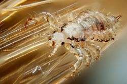
The head louse (Pediculus humanus capitis) is an obligate ectoparasite of humans that causes head lice infestation (pediculosis capitis).[1] Head lice are wingless insects spending their entire life on the human scalp and feeding exclusively on human blood.[1] Humans are the only known hosts of this specific parasite, while chimpanzees host a closely related species, Pediculus schaeffi. Other species of lice infest most orders of mammals and all orders of birds,[1] as well as other parts of the human body.
Lice differ from other hematophagic ectoparasites such as fleas in spending their entire life cycle on a host.[2] Head lice cannot fly, and their short stumpy legs render them incapable of jumping, or even walking efficiently on flat surfaces.[2]
The non-disease-carrying head louse differs from the related disease-carrying body louse (Pediculus humanus humanus) in preferring to attach eggs to scalp hair rather than to clothing. The two subspecies are morphologically almost identical but do not normally interbreed, although they will do so in laboratory conditions. From genetic studies, they are thought to have diverged as subspecies about 30,000–110,000 years ago, when many humans began to wear a significant amount of clothing.[3][4] A much more distantly related species of hair-clinging louse, the pubic or crab louse (Pthirus pubis), also infests humans. It is visually different from the other two species and is much closer in appearance to the lice which infest other primates.[5] Lice infestation of any part of the body is known as pediculosis.[6]
Head lice (especially in children) have been, and still are, subject to various eradication campaigns. Unlike body lice, head lice are not the vectors of any known diseases. Head lice are not easily contagious, but it is possible in adults. Exposure or association to a contaminated individual for more than 10 minutes results in an 85% chance in becoming exposed. If you are contaminated with head lice for more than 24 hours without using lice shampoo, it is almost certain you are infested and need to quarantine and remove yourself from your living space. Except for rare secondary infections that result from scratching at bites, head lice are harmless, and they have been regarded by some as essentially a cosmetic rather than a medical problem. It has even been suggested that head lice infestations might be beneficial in helping to foster a natural immune response against lice which helps humans in defense against the far more dangerous body louse, which is capable of transmission of dangerous diseases.[7]
Adult morphology
Like other insects of the suborder Anoplura, adult head lice are small (2.5–3 mm long), dorso-ventrally flattened (see anatomical terms of location), and entirely wingless.[8] The thoracic segments are fused, but otherwise distinct from the head and abdomen, the latter being composed of seven visible segments.[9] Head lice are grey in general, but their precise color varies according to the environment in which they were raised.[9] After feeding, consumed blood causes the louse body to take on a reddish color.[9]
Head


One pair of antennae, each with five segments, protrude from the insect's head. Head lice also have one pair of eyes. Eyes are present in all species within Pediculidae (the family of which the head louse is a member) but are reduced or absent in most other members of the Anoplura suborder.[8] Like other members of Anoplura, head lice mouth parts are highly adapted for piercing skin and sucking blood.[8] These mouth parts are retracted into the insect's head except during feeding.[9][10]
Thorax

Six legs project from the fused segments of the thorax.[9] As is typical in Anoplura, these legs are short and terminate with a single claw and opposing "thumb".[9] Between its claw and thumb, the louse grasps the hair of its host.[9] With their short legs and large claws, lice are well adapted to clinging to the hair of their host. These adaptations leave them incapable of jumping, or even walking efficiently on flat surfaces. Lice can climb up strands of hair very quickly, allowing them to move quickly and reach another host.[2]
Abdomen
There are seven visible segments of the louse abdomen.[9] The first six segments each have a pair of spiracles through which the insect breathes.[9] The last segment contains the anus and (separately) the genitalia.[9]
Sex differences
In male lice, the front two legs are slightly larger than the other four. This specialized pair of legs is used for holding the female during copulation. Males are slightly smaller than females and are characterized by a pointed end of the abdomen and a well-developed genital apparatus visible inside the abdomen. Females are characterized by two gonopods in the shape of a W at the end of their abdomen.
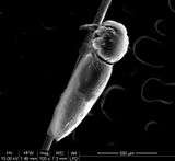
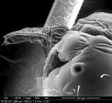
Louse eggs

Like most insects, head lice are oviparous. Females lay about 3–4 eggs per day. Louse eggs are attached near the base of a host hair shaft.[11][12] Egg-laying behavior is temperature dependent and likely seeks to place the egg in a location that will be conducive to proper embyro development (which is, in turn, temperature dependent). In cool climates, eggs are generally laid within 3–5 mm of the scalp surface.[11][12] In warm climates, and especially the tropics, eggs may be laid 6 inches (15 cm) or more down the hair shaft.[13]
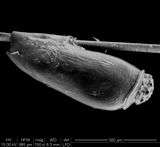
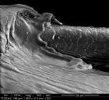
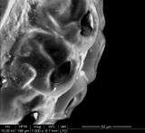
To attach an egg, the adult female secretes a glue from her reproductive organ. This glue quickly hardens into a "nit sheath" that covers the hair shaft and large parts of the egg except for the operculum, a cap through which the embryo breathes.[12] The glue was previously thought to be chitin-based, but more recent studies have shown it to be made of proteins similar to hair keratin.[12]
Each egg is oval-shaped and about 0.8 mm in length.[12] They are bright, transparent, tan to coffee-colored so long as they contain an embryo but appear white after hatching.[12][13] Typically, a hatching time of six to nine days after oviposition is cited by authors.[11][14]
After hatching, the louse nymph leaves behind its egg shell (usually known as nit), still attached to the hair shaft. The empty egg shell remains in place until physically removed by abrasion or the host, or until it slowly disintegrates, which may take 6 or more months.[14]
| SEM images of a hair louse egg | ||||||||||
|---|---|---|---|---|---|---|---|---|---|---|
|
Nits
The term nit refers to an egg without embryo or a dead egg.[15] With respect to eggs, this rather broad definition includes the following:[16] Accordingly, on the head of an infested individual the following eggs could be found:
- Viable eggs that will eventually hatch
- Remnants of already-hatched eggs (nits)
- Nonviable eggs (dead embryo) that will never hatch
This has produced some confusion in, for example, school policy (see The "no-nit" policy) because, of the three items listed above, only eggs containing viable embryos have the potential to infest or reinfest a host.[17] Some authors have reacted to this confusion by restricting the definition of nit to describe only a hatched or nonviable egg:

In many languages the terms used for the hatched eggs, which were obvious for all to see, have subsequently become applied to the embryonated eggs that are difficult to detect. Thus the term "nit" in English is often used for both. However, in recent years my colleagues and I have felt the need for some simple means of distinguishing between the two without laborious qualification. We have, therefore, come to reserve the term "nit" for the hatched and empty egg shell and refer to the developing embryonated egg as an "egg".— Ian F. Burgess (1995)[14]
The empty eggshell, termed a nit...— J. W. Maunder (1983)[2]
...nits (dead eggs or empty egg cases)...— Kosta Y. Mumcuoglu and others (2006)[18]
Others have retained the broad definition while simultaneously attempting to clarify its relevance to infestation:
In the United States the term "nit" refers to any egg regardless of its viability.— Terri Lynn Meinking (1999)[13]
Because nits are simply egg casings that can contain a developing embryo or be empty shells, not all nits are infective.— L. Keoki Williams and others (2001)[11]
Development and nymphs

Head lice, like other insects of the order Phthiraptera, are hemimetabolous.[1][10] Newly hatched nymphs will moult three times before reaching the sexually-mature adult stage.[1] Thus, mobile head lice populations contain members of up to four developmental stages: three nymphal instars, and the adult (imago).[1] Metamorphosis during head lice development is subtle. The only visible differences between different instars and the adult, other than size, is the relative length of the abdomen, which increases with each molt.[1] Aside from reproduction, nymph behavior is similar to the adult. Nymphs feed only on human blood (hematophagia), and cannot survive long away from a host.[1]
The time required for head lice to complete their nymph development to the imago depends on feeding conditions. At minimum, eight to nine days is required for lice having continuous access to a human host.[1] This experimental condition is most representative of head lice conditions in the wild. Experimental conditions where the nymph has more limited access to blood produces more prolonged development, ranging from 12 to 24 days.[1]
Nymph mortality in captivity is high—about 38%—especially within the first two days of life.[1] In the wild, mortality may instead be highest in the third instar.[1] Nymph hazards are numerous. Failure to completely hatch from the egg is invariably fatal and may be dependent on the humidity of the egg's environment.[1] Death during molting can also occur, although it is reportedly uncommon.[1] During feeding, the nymph gut can rupture, dispersing the host's blood throughout the insect. This results in death within a day or two.[1] It is unclear if the high mortality recorded under experimental conditions is representative of conditions in the wild.[1]
Reproduction and lifespan

Adult head lice reproduce sexually, and copulation is necessary for the female to produce fertile eggs. Parthenogenesis, the production of viable offspring by virgin females, does not occur in Pediculus humanus.[1] Pairing can begin within the first 10 hours of adult life.[1] After 24 hours, adult lice copulate frequently, with mating occurring during any period of the night or day.[1][19] Mating attachment frequently lasts more than an hour.[19] Young males can successfully pair with older females, and vice versa.[1]
Experiments with Pediculus humanus humanus (body lice) emphasize the attendant hazards of lice copulation. A single young female confined with six or more males will die in a few days, having laid very few eggs.[1] Similarly, death of a virgin female was reported after admitting a male to her confinement.[19] The female laid only one egg after mating, and her entire body was tinged with red—a condition attributed to rupture of the alimentary canal during the sexual act.[19] Old females frequently die following, if not during, intercourse.[19]
A single louse has a thirty-day life cycle beginning from the moment the nit is laid until the adult louse dies.[20]
Factors affecting infestation
The number of children per family, the sharing of beds and closets, hair washing habits, local customs and social contacts, healthcare in a particular area (e.g. school) and socioeconomic status were found to be significant factors in head louse infestation. Girls are two to four times more frequently infested than boys. Children between 4 and 14 years of age are the most frequently infested group.[20]
Behaviour
Feeding
All stages are blood-feeders and bite the skin four to five times daily to feed. They inject saliva which contains an anti-coagulant and suck blood. The digested blood is excreted as dark red frass.[21]
Position on host
Although any part of the scalp may be colonized, lice favor the nape of the neck and the area behind the ears, where the eggs are usually laid. Head lice are repelled by light and will move towards shadows or dark-coloured objects in their vicinity.[19][22]
Transmission
Lice have no wings or powerful legs for jumping, so they move by using their claw-like legs to transfer from hair to hair.[21] Normally head lice infest a new host only by close contact between individuals, making social contacts among children and parent-child interactions more likely routes of infestation than shared combs, hats, brushes, towels, clothing, beds or closets. Head-to-head contact is by far the most common route of lice transmission.[23]
Distribution
About 6–12 million people, mainly children, are treated annually for head lice in the United States alone. High levels of louse infestations have also been reported from all over the world, including Australia, Denmark, France, Ireland, Israel and Sweden.[17][24] Head lice can live off the head, for example on soft furnishings such as pillow cases, on hairbrushes, or on coat hoods for up to 48 hours.
Archaeogenetics
Analysis of the DNA of lice found on Peruvian mummies may indicate that some diseases (like typhus) may have passed from the New World to the Old World, instead of the other way around.[25]
Genome
The sequencing of the genome of the body louse was first proposed in the mid-2000s[26] and the annotated genome was published in 2010.[27] An analysis of the body and head louse transcriptomes revealed these two organisms are extremely similar genetically.[28]
See also
References
- 1 2 3 4 5 6 7 8 9 10 11 12 13 14 15 16 17 18 19 20 21 Buxton, Patrick A. (1947). "The biology of Pediculus humanus". The Louse; an account of the lice which infest man, their medical importance and control (2nd ed.). London: Edward Arnold. pp. 24–72.
- 1 2 3 4 Maunder, JW (1983). "The Appreciation of Lice". Proceedings of the Royal Institution of Great Britain. London. 55: 1–31.
- ↑ Kittler R, Kayser M, Stoneking M (August 2003). "Molecular evolution of Pediculus humanus and the origin of clothing". Current Biology. 13 (16): 1414–7. doi:10.1016/S0960-9822(03)00507-4. PMID 12932325.
- ↑ Stoneking, Mark (29 December 2004). "Erratum: Molecular Evolution of Pediculus humanus and the Origin of Clothing". Current Biology. 14 (24): 2309. doi:10.1016/j.cub.2004.12.024.
- ↑ Buxton, Patrick A. (1947). "The crab louse Phthirus pubis". The Louse; an account of the lice which infest man, their medical importance and control (2nd ed.). London: Edward Arnold. pp. 136–141.
- ↑ "pediculosis – Definition from the Merriam-Webster Online Dictionary". Retrieved 2008-04-23.
- ↑ Rozsa, L; Apari, P. (2012). "Why infest the loved ones – inherent human behaviour indicates former mutualism with head lice" (PDF). Parasitology. 139: 696–700. doi:10.1017/s0031182012000017.
- 1 2 3 Buxton, Patrick A. (1947). "The Anoplura or Sucking Lice". The Louse; an account of the lice which infest man, their medical importance and control (2nd ed.). London: Edward Arnold. pp. 1–4.
- 1 2 3 4 5 6 7 8 9 10 Buxton, Patrick A. (1947). "The Anatomy of Pediculus humanus". The Louse; an account of the lice which infest man, their medical importance and control (2nd ed.). London: Edward Arnold. pp. 5–23.
- 1 2 "Lice (Pediculosis)". The Merck Veterinary Manual. Whitehouse Station, NJ USA: Merck & Co. 2008. Retrieved 2008-10-08.
- 1 2 3 4 Williams LK, Reichert A, MacKenzie WR, Hightower AW, Blake PA (May 2001). "Lice, nits, and school policy". Pediatrics. 107 (5): 1011–5. doi:10.1542/peds.107.5.1011. PMID 11331679.
- 1 2 3 4 5 6 Burkhart CN, Burkhart CG (July 2005). "Head lice: scientific assessment of the nit sheath with clinical ramifications and therapeutic options". Journal of the American Academy of Dermatology. 53 (1): 129–33. doi:10.1016/j.jaad.2005.01.134. PMID 15965432.
- 1 2 3 Meinking, Terri Lynn (May–June 1999). "Infestations". Current Problems in Dermatology. 11 (3): 75–118. doi:10.1016/S1040-0486(99)90005-4.
- 1 2 3 Burgess, IF (1995). "Human lice and their management". Advances in parasitology. Advances in Parasitology. 36: 271–342. doi:10.1016/S0065-308X(08)60493-5. ISBN 978-0-12-031736-3. PMID 7484466.
- ↑ "nit – Definition from the Merriam-Webster Online Dictionary". Retrieved 2008-02-25.
- ↑ Pollack RJ, Kiszewski AE, Spielman A (August 2000). "Overdiagnosis and consequent mismanagement of head louse infestations in North America". The Pediatric Infectious Disease Journal. 19 (8): 689–93; discussion 694. doi:10.1097/00006454-200008000-00003. PMID 10959734.
- 1 2 Burgess, IF (2004). "Human lice and their control". Annu. Rev. Entomol. 49: 457–81. doi:10.1146/annurev.ento.49.061802.123253. PMID 14651472.
- ↑ Mumcuoglu KY, Meinking TA, Burkhart CN, Burkhart CG (August 2006). "Head louse infestations: the 'no nit' policy and its consequences". International Journal of Dermatology. 45 (8): 891–6. doi:10.1111/j.1365-4632.2006.02827.x. PMID 16911370.
- 1 2 3 4 5 6 Bacot A (1917). "Contributions to the bionomics of Pediculus humanus (vestimenti) and Pediculus capitis". Parasitology. 9 (2): 228–258. doi:10.1017/S0031182000006065.
- 1 2 Mumcuoglu KY, Miller J, Gofin R, et al. (September 1990). "Epidemiological studies on head lice infestation in Israel. I. Parasitological examination of children". International Journal of Dermatology. 29 (7): 502–6. doi:10.1111/j.1365-4362.1990.tb04845.x. PMID 2228380.
- 1 2 Weems, Jr., H. V.; Fasulo, T. R. (June 2007). "Human Lice: Body Louse, Pediculus humanus humanus Linnaeus and Head Louse, Pediculus humanus capitis De Geer (Insecta: Phthiraptera (=Anoplura): Pediculidae)". University of Florida, Institute of Food and Agricultural Sciences. Retrieved 2008-02-21.
- ↑ Nuttall, George H. F. (1919). "The biology of Pediculus humanus, Supplementary notes". Parasitology. 11 (2): 201–221. doi:10.1017/s0031182000004194.
- ↑ "NJ Head Lice | Philadelphia and South New Jersey Hair Lice". Lice Lifters New Jersey. Retrieved 2012-11-22.
- ↑ Mumcuoglu KY, Barker SC, Burgess IE, et al. (April 2007). "International guidelines for effective control of head louse infestations". Journal of Drugs in Dermatology. 6 (4): 409–14. PMID 17668538.
- ↑ Anderson, Andrea (February 8, 2008). "DNA from Peruvian Mummy Lice Reveals History". GenomeWeb Daily News. GenomeWeb LLC. Retrieved August 31, 2014.
- ↑ Pittendrigh BR, Clark JM, Johnston JS, Lee SH, Romero-Severson J, Dasch GA (November 2006). "Sequencing of a new target genome: the Pediculus humanus humanus (Phthiraptera: Pediculidae) genome project". Journal of Medical Entomology. 43 (6): 1103–11. doi:10.1603/0022-2585(2006)43[1103:SOANTG]2.0.CO;2. PMID 17162941.
- ↑ Kirkness EF, Haas BJ, Sun W, et al. (July 2010). "Genome sequences of the human body louse and its primary endosymbiont provide insights into the permanent parasitic lifestyle". Proceedings of the National Academy of Sciences of the United States of America. 107 (27): 12168–73. doi:10.1073/pnas.1003379107. PMC 2901460
 . PMID 20566863.
 - ↑ Olds BP, Coates BS, Steele LD, et al. (April 2012). "Comparison of the transcriptional profiles of head and body lice". Insect Molecular Biology. 21 (2): 257–68. doi:10.1111/j.1365-2583.2012.01132.x. PMID 22404397.
External links
| Look up nit in Wiktionary, the free dictionary. |
| Wikimedia Commons has media related to Pediculus humanus. |
| Look up head louse in Wiktionary, the free dictionary. |
- Centers for Disease Control and Prevention: Division of Parasitic Diseases
- James Cook University, Australia: Head Lice Information Sheet
- University of Nebraska: Head Lice Resources You Can Trust
- body and head lice on the UF / IFAS Featured Creatures Web site