Gregarinasina
| Gregarinasina | |
|---|---|
 | |
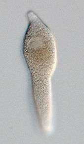
| A live specimen of a septate (or cephaline) gregarine showing the distinctive "head"-like section of the trophozoite containing the epimerite at its anterior end. Septate gregarines are intestinal parasites of arthropods. | |
| Scientific classification | |
| Domain: | Eukaryota |
| (unranked): | SAR |
| (unranked): | Alveolata |
| Phylum: | Apicomplexa |
| Class: | Conoidasida |
| Subclass: | Gregarinasina |
| Orders | |
| Synonyms | |
| |
The gregarines are a group of Apicomplexan protozoa, classified as the Gregarinasina[1] or Gregarinia. The large (roughly half a millimeter) parasites inhabit the intestines of a large number of invertebrates. They are not found in any vertebrates. However, gregarines are closely related to both Toxoplasma and Plasmodium, which cause toxoplasmosis and malaria, respectively. Both protists use protein complexes similar to those that are formed by the gregarines for gliding motility and invading target cells.[2][3] This makes them an excellent model for studying gliding motility with the goal of developing treatment options for toxoplasmosis and malaria.
Lifecycle
Gregarines occur in both aquatic and terrestrial environments. Although they are usually transmitted by the orofaecal route, some are transmitted with the host's gametes during copulation (Monocystis).
In all species, four or more sporozoites (the precise number depends on the species) equipped with an apical complex escape from the oocysts, a process called excystation, find their way to the appropriate body cavity, and penetrate host cells in their immediate environment. The sporozoites emerge within the host cell, begin to feed, and develop into larger trophozoites. In some species, the sporozoites and trophozoites are capable of asexual replication — a process called schizogony or merogony. Most species, however, appear to lack schizogony in their lifecycles.
In all species, two mature trophozoites eventually pair up in a process known as syzygy and develop into gamonts. During syzygy, gamont orientation differs between species (side-to-side, head-to-tail). A gametocyst wall forms around each pair of gamonts which then begin to divide into hundreds of gametes. Zygotes are produced by the fusion of two gametes and these in turn become surrounded by an oocyst wall. Within the oocyst, meiosis occurs, yielding the sporozoites. Hundreds of oocysts accumulate within each gametocyst and these are released via host's faeces or via host death and decay.
Gregarines have been reported to infect over 3000 invertebrate species.[4]
Taxonomy
The gregarines were recognised as a taxon by Grasse in 1953.[5] The three orders into which they are currently divided were created by Levine et al in 1980.
Currently, about 250 genera and 1650 species are known in this taxon. They are divided into three orders based on habitat, host range, and trophozoite morphology.[6]
Most species have monoxenous life cycles involving a single invertebrate host. In the life cycle is an extracellular feeding stage known as the trophozoite.
Main divisions
Archigregarines are found only in marine habitats. They possess intestinal trophozoites similar in morphology to the infective sporozoites. Phylogenetic analysis suggests this group is paraphyletic and will need division. Generally, four zoites are in each spore in this group.
Eugregarines are found in marine, freshwater, and terrestrial habitats. These species possess large trophozoites that are significantly different in morphology and behavior from the sporozoites. This taxon contains most of the known gregarine species. The intestinal eugregarines are separated into septate — suborder Septatorina — and aseptate — suborder Aseptatorina — depending on whether the trophozoite is superficially divided by a transverse septum. The aseptate species are mostly marine gregarines.
Urosporidians are aseptate eugregarines that infect the coelomic spaces of marine hosts. Unusually, they tend to lack attachment structures and form gamont pairs that pulsate freely within the coelomic fluid.
Monocystids are aseptate eugregarines that infect the reproductive vesicles of terrestrial annelids. These latter species tend to branch closely with neogregarines and may need to be reclassified. Generally, eight zoites are in each spore in this group.
Neogregarines are found only in terrestrial hosts. These species have reduced trophozoites and tend to infect tissues other than the intestine. Usually, eight zoites are in each spore in this group.
The eugregarines and neogregarines differ in a number of respects. The neogregarines are in general more pathogenic to their hosts. The eugregarines multiply by sporogony and gametogony, while the neogregarines have an additional schizogenic stage — merogony — within their hosts. Merogony may be intracellular or extracellular depending on the species.
DNA studies suggest the archigregarines are ancestral to the others.[7]
Proposed revision
Cavalier-Smith has proposed a significant revision of this taxon.[8]
He has suggested a new class - Gregarinomorphea - which includes Orthogregarinia, Cryptosporidiidae and Rhytidocystidae. The Orthogregarinia with two new orders Arthrogregarida and Vermigregarida was created for the gregarines most closely related to Cryptosporidium.
A second class - Paragregarea - was created for the archigregarines, Stenophorida and a new order - Velocida which itself was created for Urosporoidea superfam. n. and Veloxidium.
A third order was created - Squirmida - for Filipodium and Platyproteum.
Notes
There are several genera of gregarines that are currently not classified: Acuta, Cephalolobus, Gregarina, Levinea, Menospora, Nematocystis, Nematopsis, Steinina and Trichorhynchus.
Characteristics
- Meiosis occurs in all species.
- Monoxenous — only one host occurs in lifecycle for almost all species.
- Mitochondria have tubular cristae and are often distributed near the cell periphery.
- Apical complex occurs in the sporozoite stage, but is lost in the trophozoite stage in eugregarines and neogregarines.
- Trophozoites have a large and conspicuous nucleus and nucleolus.
- They inhabit extracellular body cavities of invertebrates such as the intestines, coeloms, and reproductive vesicles.
- Attachment to host occurs by a mucron (aseptate gregarines) or an epimerite (septate gregarines); some gregarines (urosporidians) float freely within extracellular body cavities (coelom).
The parasites are relatively large spindle-shaped cells, compared to other apicomplexans and eukaryotes in general (some species are > 850 µm in length). Most gregarines have longitudinal epicytic folds (bundles of microtubules beneath the cell surface with nematode like bending behaviour): crenulations are instead found in the urosporidians.
Molecular biology
The gregarines are able to move and change direction along a surface through gliding motility without the use of cilia, flagella, or lamellipodia.[9] This is accomplished through the use of an actin and myosin complex.[10] The complexes require an actin cytoskeleton to perform their gliding motions.[11] In the proposed ‘capping’ model, an uncharacterized protein complex moves rearward, moving the parasites forward.[12]
History
The gregarines are among the oldest known parasites having been described by the physician Francesco Redi in 1684.[13]
The first formal description was made by Dufour in 1828. He created the genus Gregarina and described Gregarina ovata from Folficula aricularia. He considered them to be parasitic worms. Koelliker recognised them as protozoa in 1848.
References
- ↑ Carreno RA, Martin DS, Barta JR (November 1999). "Cryptosporidium is more closely related to the gregarines than to coccidia as shown by phylogenetic analysis of apicomplexan parasites inferred using small-subunit ribosomal RNA gene sequences". Parasitol. Res. 85 (11): 899–904. doi:10.1007/s004360050655. PMID 10540950.
- ↑ Ménard R (February 2001). "Gliding motility and cell invasion by Apicomplexa: insights from the Plasmodium sporozoite". Cell. Microbiol. 3 (2): 63–73. doi:10.1046/j.1462-5822.2001.00097.x. PMID 11207621.
- ↑ Meissner M, Schlüter D, Soldati D (October 2002). "Role of Toxoplasma gondii myosin A in powering parasite gliding and host cell invasion". Science. 298 (5594): 837–40. doi:10.1126/science.1074553. PMID 12399593.
- ↑ Alarcón M E., Huang C-G, Tsai Y-S, Chen W-J, Kumar A (2011) Life cycle and morphology of Steinina ctenocephali (Ross 1909) comb. nov. (Eugregarinorida: Actinocephalidae), a gregarine of Ctenocephalides felis (Siphonaptera: Pulicidae) in Taiwan. Zoological Studies 50(6): 763-772
- ↑ Grassé, P.P.; Caullery, M.C. (1953). Traité de zoologie: anatomie, systématique, biologie. Tome I, Fasc. II, Protozaires, rhizopodes, Actinopodes, Sporozoaires, Cnidosporidies. Paris: Masson et Cie. OCLC 642231286.
- ↑ Perkins FO, Barta JR, Clopton RE, Peirce MA, Upton SJ (2000). "Phylum Apicomplexa". In Lee JJ, Leedale GF, Bradbury P. An Illustrated guide to the Protozoa: organisms traditionally referred to as protozoa, or newly discovered groups. 1 (2nd ed.). Society of Protozoologists. pp. 190–369. ISBN 1891276220. OCLC 704052757.
- ↑ Leander BS (February 2008). "Marine gregarines: evolutionary prelude to the apicomplexan radiation?". Trends Parasitol. 24 (2): 60–7. doi:10.1016/j.pt.2007.11.005. PMID 18226585.
- ↑ Cavalier-Smith T (2014) Gregarine site-heterogeneous 18S rDNA trees, revision of gregarine higher classification, and the evolutionary diversification of Sporozoa. European Journal of Protistology 50 (5) 472–495
- ↑ Walker MH, Mackenzie C, Bainbridge SP, Orme C (November 1979). "A study of the structure and gliding movement of Gregarina garnhami". J Protozool. 26: 566–574. doi:10.1111/j.1550-7408.1979.tb04197.x.
- ↑ Heintzelman MB (June 2004). "Actin and myosin in Gregarina polymorpha". Cell Motil. Cytoskeleton. 58 (2): 83–95. doi:10.1002/cm.10178. PMID 15083530.
- ↑ Mitchison TJ, Cramer LP (February 1996). "Actin-based cell motility and cell locomotion". Cell. 84 (3): 371–9. doi:10.1016/s0092-8674(00)81281-7. PMID 8608590.
- ↑ Sibley LD, Hâkansson S, Carruthers VB (January 1998). "Gliding motility: an efficient mechanism for cell penetration". Curr. Biol. 8 (1): R12–4. doi:10.1016/S0960-9822(98)70008-9. PMID 9427622.
- ↑ Redi, F (1684). Osservazioni intorno agli animali viventi, che si trovano negli animali viventi. Firenze: P. Matini. OCLC 8705660.
Bibliography
- Desportes, I. & Schrével, J. (2013). Treatise on Zoology - Anatomy, Taxonomy, Biology. The Gregarines. 2 vols. Leiden, Netherlands: Brill, .
External links
- Tree of Life Gregarina
- Gregarina Movies
- "Gregarinasina". NCBI Taxonomy Browser. 35086.