Glycogen synthase
| glycogen (starch) synthase | |||||||||
|---|---|---|---|---|---|---|---|---|---|
 | |||||||||
| Identifiers | |||||||||
| EC number | 2.4.1.11 | ||||||||
| CAS number | 9014-56-6 | ||||||||
| Databases | |||||||||
| IntEnz | IntEnz view | ||||||||
| BRENDA | BRENDA entry | ||||||||
| ExPASy | NiceZyme view | ||||||||
| KEGG | KEGG entry | ||||||||
| MetaCyc | metabolic pathway | ||||||||
| PRIAM | profile | ||||||||
| PDB structures | RCSB PDB PDBe PDBsum | ||||||||
| Gene Ontology | AmiGO / EGO | ||||||||
| |||||||||
Glycogen synthase (UDP-glucose-glycogen glucosyltransferase) is a key enzyme in glycogenesis, the conversion of glucose into glycogen. It is a glycosyltransferase (EC 2.4.1.11) that catalyses the reaction of UDP-glucose and (1,4-α-D-glucosyl)n to yield UDP and (1,4-α-D-glucosyl)n+1. In other words, this enzyme combines excess glucose residues one by one into a polymeric chain for storage as glycogen.
Glycogen synthase concentration is highest in the bloodstream 30 to 60 minutes following intense exercise.[2]
Structure
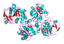
Much research has been done on glycogen degradation through studying the structure and function of glycogen phosphorylase, the key regulatory enzyme of glycogen degradation.[3] On the other hand, much less is known about the structure of glycogen synthase, the key regulatory enzyme of glycogen synthesis. The crystal structure of glycogen synthase from Agrobacterium tumefaciens, however, has been determined at 2.3 A resolution.[4] In its asymmetric form, glycogen synthase is found as a dimer, whose monomers are composed of two Rossmann-fold domains. This structural property, among others, is shared with related enzymes, such as glycogen phosphorylase and other glycosyltransferases of the GT-B superfamily.[5] Nonetheless, a more recent characterization of the Saccharomyces cerevisiae (yeast) glycogen synthase crystal structure reveals that the dimers may actually interact to form a tetramer. Specifically, The inter-subunit interactions are mediated by the α15/16 helix pairs, forming allosteric sites between subunits in one combination of dimers and active sites between subunits in the other combination of dimers. Since the structure of eukaryotic glycogen synthase is highly conserved among species, glycogen synthase likely forms a tetramer in humans as well.[6]
Glycogen synthase can be classified in two general protein families. The first family (GT3), which is from mammals and yeast, is approximately 80 kDa, uses UDP-glucose as a sugar donor, and is regulated by phosphorylation and ligand binding.[7] The second family (GT5), which is from bacteria and plants, is approximately 50 kDA, uses ADP-glucose as a sugar donor, and is unregulated.[8]
Mechanism
Although the catalytic mechanisms used by glycogen synthase are not well known, structural similarities to glycogen phosphorylase at the catalytic and substrate binding site suggest that the mechanism for synthesis is similar in glycogen synthase and glycogen phosphorylase.[4]
Function
Glycogen synthase catalyzes the conversion of the glucosyl (Glc) moiety of uridine diphosphate glucose (UDP-Glc) into glucose to be incorporated into glycogen via an α(1→4) glycosidic bond. However, since glycogen synthase requires an oligosaccharide primer as a glucose acceptor, it relies on glycogenin to initiate de novo glycogen synthesis.[6]
In a recent study of transgenic mice, an overexpression of glycogen synthase[9] and an overexpression of phosphatase[10] both resulted in excess glycogen storage levels. This suggests that glycogen synthase plays an important biological role in regulating glycogen/glucose levels and is activated by dephosphorylation.
Isozymes
In humans, there are two paralogous isozymes of glycogen synthase:
| isozyme | tissue distribution | gene |
|---|---|---|
| glycogen synthase 1 containing carbon elements | muscle and other tissues | GYS1[11] |
| glycogen synthase 2 | liver | GYS2[12] |
The liver enzyme expression is restricted to the liver, whereas the muscle enzyme is widely expressed. Liver glycogen serves as a storage pool to maintain the blood glucose level during fasting, whereas muscle glycogen synthesis accounts for disposal of up to 90% of ingested glucose. The role of muscle glycogen is as a reserve to provide energy during bursts of activity.[13]
Meanwhile, the muscle isozyme plays a major role in the cellular response to long-term adaptation to hypoxia. Notably, hypoxia only induces expression of the muscle isozyme and not the liver isozyme. However, muscle-specific glycogen synthase activation may lead to excessive accumulation of glycogen, leading to damage in the heart and central nervous system following ischemic insults.[14]
|
| ||||||||||||||||||||||||||||||||||||||||||||
Regulation
The reaction is highly regulated by allosteric effectors such as glucose-6-phosphate (which allows glycogen synthase to operate as a glucose-6-phosphate sensor), by phosphorylation reactions (catalyzed by GSK3, see below), and indirectly triggered by the hormone insulin, which is secreted by the pancreas in response to elevated blood glucose levels. Phosphorylation of glycogen synthase decreases its activity. The enzyme also cleaves the ester bond between the C1 position of glucose and the pyrophosphate of UDP itself.
The control of glycogen synthase is a key step in regulating glycogen metabolism and glucose storage. Glycogen synthase is directly regulated by glycogen synthase kinase 3 (GSK-3), AMPK, protein kinase A (PKA), and casein kinase 2 (CK2). Each of these protein kinases lead to phosphorylated and catalytically inactive glycogen synthase. The phosphorylation sites of glycogen synthase are summarized below.
| Name | Phosphorylation Site | Kinase | Reference(s) |
|---|---|---|---|
| Site 1a | PKA | ,[15][16] | |
| Site 1b | PKA | ,[15][16] | |
| Site 2 | Serine 7 | AMPK | ,[17][18] |
| Site 2a | Serine 10 | CK2 | |
| Site 3a | Serine 641 | GSK3 | [19] |
| Site 3b | Serine 645 | GSK3 | [19] |
| Site 3c | Serine 649 | GSK3 | [19] |
| Site 3d | Serine 653 | GSK3 | [19] |
| Site 4 | Serine 727 |
For enzymes in the GT3 family, these regulatory kinases inactivate glycogen synthase by phosphorylating it at the N-terminal of the 25th residue and the C-terminal of the 120th residue.[4] Glycogen synthase is also regulated by protein phosphatase 1 (PP1), which activates glycogen synthase via dephosphorylation.[20] PP1 is targeted to the glycogen pellet by four targeting subunits, GM, GL, PTG and R6. These regulatory enzymes are regulated by insulin and glucagon signaling pathways.
Clinical significance
Mutations in the GYS1 gene are associated with glycogen storage disease type 0.[21] In humans, defects in the tight control of glucose uptake and utilization are also associated with diabetes and hyperglycemia. Patients with type 2 diabetes normally exhibit low glycogen storage levels because of impairments in insulin-stimulated glycogen synthesis and suppression of glycogenolysis. Insulin stimulates glycogen synthase by inhibiting glycogen synthase kinases or/and activating protein phosphatase 1 (PP1) among other mechanisms.[20]
Model organisms
| Characteristic | Phenotype |
|---|---|
| Homozygote viability | Normal |
| Fertility | Normal |
| Body weight | Normal |
| Anxiety | Normal |
| Neurological assessment | Normal |
| Grip strength | Normal |
| Hot plate | Normal |
| Dysmorphology | Normal |
| Indirect calorimetry | Normal |
| Glucose tolerance test | Abnormal |
| Auditory brainstem response | Normal |
| DEXA | Normal |
| Radiography | Normal |
| Body temperature | Normal |
| Eye morphology | Normal |
| Clinical chemistry | Abnormal[22] |
| Plasma immunoglobulins | Normal |
| Haematology | Normal |
| Peripheral blood lymphocytes | Normal |
| Micronucleus test | Normal |
| Heart weight | Normal |
| Skin Histopathology | Normal |
| Brain histopathology | Normal |
| Eye Histopathology | Normal |
| Salmonella infection | Normal[23] |
| Citrobacter infection | Normal[24] |
| All tests and analysis from[25][26] |
Model organisms have been used in the study of GYS2 function. A conditional knockout mouse line, called Gys2tm1a(KOMP)Wtsi[27][28] was generated as part of the International Knockout Mouse Consortium program — a high-throughput mutagenesis project to generate and distribute animal models of disease to interested scientists — at the Wellcome Trust Sanger Institute.[29][30][31] Male and female animals underwent a standardized phenotypic screen to determine the effects of deletion.[25][32] Twenty six tests were carried out and two significant phenotypes were reported. Homozygous mutant male adults displayed impaired glucose tolerance, whereas females had a significant decrease in circulating glucose levels as determined by clinical chemistry.[25]
References
- ↑ PDB: 1RZU; Buschazzio A, Ugalde JE, Guerin ME, Shepard W, Ugalde RA, Alzari PM (August 2004). "Crystal structure of glycogen synthase: homologous enzymes catalyze glycogen synthesis and degradation". EMBO J. 23 (16): 3196–3205. doi:10.1038/sj.emboj.7600324. PMC 514502
 . PMID 15272305.; rendered using PyMOL.
 - ↑ Jentjens R, Jeukendrup A (2003). "Determinants of post-exercise glycogen synthesis during short-term recovery". Sports Med. 33 (2): 117–44. doi:10.2165/00007256-200333020-00004. PMID 12617691.
- ↑ Buchbinder JL, Rath VL, Fletterick RJ (2001). "Structural relationships among regulated and unregulated phosphorylases". Annu Rev Biophys Biomol Struct. 30 (1): 191–209. doi:10.1146/annurev.biophys.30.1.191. PMID 11340058.
- 1 2 3 Buschiazzo A, Ugalde JE, Guerin ME, Shepard W, Ugalde RA, Alzari PM (2004). "Crystal structure of glycogen synthase: homologous enzymes catalyze glycogen synthesis and degradation.". EMBO J. 23 (16): 3195–205. doi:10.1038/sj.emboj.7600324. PMC 514502. PMID 15272305.
- ↑ Coutinho PM, Deleury E, Davies GJ, Henrissat B (2003). "An evolving hierarchical family classification for glycosyltransferases". J. Mol. Biol. 328 (2): 307–17. doi:10.1016/S0022-2836(03)00307-3. PMID 12691742.
- 1 2 Palm, DC; Rohwer, JM; Hofmeyr, JH (January 2013). "Regulation of glycogen synthase from mammalian skeletal muscle--a unifying view of allosteric and covalent regulation.". The FEBS Journal. 280 (1): 2–27. doi:10.1111/febs.12059. PMID 23134486.
- ↑ Roach PJ (2002). "Glycogen and its Metabolism". Curr Mol Med. 2 (2): 101–20. doi:10.2174/1566524024605761. PMID 11949930.
- ↑ Ball SG, Morell MK (2003). "From bacterial glycogen to starch: understanding the biogenesis of the plant starch granule". Annu Rev Plant Biol. 54 (1): 207–33. doi:10.1146/annurev.arplant.54.031902.134927. PMID 14502990.
- ↑ Azpiazu I, Manchester J, Skurat AV, Roach PJ, Lawrence JC Jr (2000). "Control of glycogen synthesis is shared between glucose transport and glycogen synthase in skeletal muscle fibers". Am J Physiol Endocrinol Metab. 278 (2): E234–43. PMID 10662707.
- ↑ Aschenbach WG, Suzuki Y, Breeden K, Prats C, Hirshman MF, Dufresne SD, Sakamoto K, Vilardo PG, Steele M, Kim JH, Jing SL, Goodyear LJ, DePaoli-Roach AA (2001). "The muscle-specific protein phosphatase PP1G/R(GL)(G(M))is essential for activation of glycogen synthase by exercise". J Biol Chem. 276 (43): 39959–67. doi:10.1074/jbc.M105518200. PMID 11522787.
- ↑ Browner MF, Nakano K, Bang AG, Fletterick RJ (March 1989). "Human muscle glycogen synthase cDNA sequence: a negatively charged protein with an asymmetric charge distribution". Proceedings of the National Academy of Sciences of the United States of America. 86 (5): 1443–7. doi:10.1073/pnas.86.5.1443. PMC 286712. PMID 2493642.
- ↑ Westphal SA, Nuttall FQ (February 1992). "Comparative characterization of human and rat liver glycogen synthase". Archives of Biochemistry and Biophysics. 292 (2): 479–86. doi:10.1016/0003-9861(92)90019-S. PMID 1731614.
- ↑ Kollberg G, Tulinius M, Gilljam T, Ostman-Smith I, Forsander G, Jotorp P, Oldfors A, Holme E (October 2007). "Cardiomyopathy and exercise intolerance in muscle glycogen storage disease 0". The New England Journal of Medicine. 357 (15): 1507–14. doi:10.1056/NEJMoa066691. PMID 17928598.
- ↑ Pescador, N; Villar, D; Cifuentes, D; Garcia-Rocha, M; Ortiz-Barahona, A; Vazquez, S; Ordoñez, A; Cuevas, Y; Saez-Morales, D; Garcia-Bermejo, ML; Landazuri, MO; Guinovart, J; del Peso, L (12 March 2010). "Hypoxia promotes glycogen accumulation through hypoxia inducible factor (HIF)-mediated induction of glycogen synthase 1.". PLOS ONE. 5 (3): e9644. doi:10.1371/journal.pone.0009644. PMC 2837373. PMID 20300197.
- 1 2 Huang TS, Krebs EG (April 1977). "Amino acid sequence of a phosphorylation site in skeletal muscle glycogen synthetase". Biochem. Biophys. Res. Commun. 75 (3): 643–50. doi:10.1016/0006-291X(77)91521-2. PMID 405007.
- 1 2 Proud CG, Rylatt DB, Yeaman SJ, Cohen P (August 1977). "Amino acid sequences at the two sites on glycogen synthetase phosphorylated by cyclic AMP-dependent protein kinase and their dephosphorylation by protein phosphatase-III". FEBS Lett. 80 (2): 435–42. doi:10.1016/0014-5793(77)80493-6. PMID 196939.
- ↑ Rylatt DB, Cohen P (February 1979). "Amino acid sequence at the site on rabbit skeletal muscle glycogen synthase phosphorylated by the endogenous glycogen synthase kinase-2 activity". FEBS Lett. 98 (1): 71–5. doi:10.1016/0014-5793(79)80154-4. PMID 107044.
- ↑ Embi N, Parker PJ, Cohen P (April 1981). "A reinvestigation of the phosphorylation of rabbit skeletal-muscle glycogen synthase by cyclic-AMP-dependent protein kinase. Identification of the third site of phosphorylation as serine-7". Eur. J. Biochem. 115 (2): 405–13. doi:10.1111/j.1432-1033.1981.tb05252.x. PMID 6263629.
- 1 2 3 4 Rylatt DB, Aitken A, Bilham T, Condon GD, Embi N, Cohen P (June 1980). "Glycogen synthase from rabbit skeletal muscle. Amino acid sequence at the sites phosphorylated by glycogen synthase kinase-3, and extension of the N-terminal sequence containing the site phosphorylated by phosphorylase kinase". Eur. J. Biochem. 107 (2): 529–37. doi:10.1111/j.1432-1033.1980.tb06060.x. PMID 6772446.
- 1 2 Saltiel AR (2001). "New perspectives into the molecular pathogenesis and treatment of type 2 diabetes". Cell. 104 (4): 517–29. doi:10.1016/S0092-8674(01)00239-2. PMID 11239409.
- ↑ Orho M, Bosshard NU, Buist NR, Gitzelmann R, Aynsley-Green A, Blümel P, Gannon MC, Nuttall FQ, Groop LC (August 1998). "Mutations in the liver glycogen synthase gene in children with hypoglycemia due to glycogen storage disease type 0". The Journal of Clinical Investigation. 102 (3): 507–15. doi:10.1172/JCI2890. PMC 508911. PMID 9691087.
- ↑ "Clinical chemistry data for Gys2". Wellcome Trust Sanger Institute.
- ↑ "Salmonella infection data for Gys2". Wellcome Trust Sanger Institute.
- ↑ "Citrobacter infection data for Gys2". Wellcome Trust Sanger Institute.
- 1 2 3 Gerdin AK (2010). "The Sanger Mouse Genetics Programme: High throughput characterisation of knockout mice". Acta Ophthalmologica. 88 (S248). doi:10.1111/j.1755-3768.2010.4142.x.
- ↑ Mouse Resources Portal, Wellcome Trust Sanger Institute.
- ↑ "International Knockout Mouse Consortium".
- ↑ "Mouse Genome Informatics".
- ↑ Skarnes, W. C.; Rosen, B.; West, A. P.; Koutsourakis, M.; Bushell, W.; Iyer, V.; Mujica, A. O.; Thomas, M.; Harrow, J.; Cox, T.; Jackson, D.; Severin, J.; Biggs, P.; Fu, J.; Nefedov, M.; De Jong, P. J.; Stewart, A. F.; Bradley, A. (2011). "A conditional knockout resource for the genome-wide study of mouse gene function". Nature. 474 (7351): 337–342. doi:10.1038/nature10163. PMC 3572410. PMID 21677750.
- ↑ Dolgin E (June 2011). "Mouse library set to be knockout". Nature. 474 (7351): 262–3. doi:10.1038/474262a. PMID 21677718.
- ↑ Collins FS, Rossant J, Wurst W (January 2007). "A mouse for all reasons". Cell. 128 (1): 9–13. doi:10.1016/j.cell.2006.12.018. PMID 17218247.
- ↑ van der Weyden L, White JK, Adams DJ, Logan DW (2011). "The mouse genetics toolkit: revealing function and mechanism.". Genome Biol. 12 (6): 224. doi:10.1186/gb-2011-12-6-224. PMC 3218837. PMID 21722353.
External links
- Glycogen Synthase at the US National Library of Medicine Medical Subject Headings (MeSH)
- Newcastle University Centre for Cancer Education (October 9, 1997). "Glycogen synthetase". Retrieved 2007-11-05.