Gastroschisis
| Gastroschisis | |
|---|---|
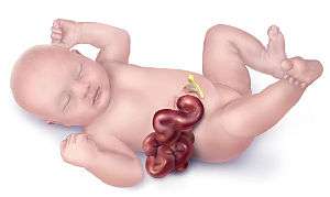 | |
| Illustration of a child with gastroschisis | |
| Classification and external resources | |
| Specialty | medical genetics |
| ICD-10 | Q79.3 |
| ICD-9-CM | 756.73 |
| OMIM | 230750 |
| DiseasesDB | 31155 |
| MedlinePlus | 000992 |
| eMedicine | ped/1642 radio/303 |
| MeSH | D020139 |

Gastroschisis represents a congenital defect characterised by a defect in the anterior abdominal wall through which the abdominal contents freely protrude. There is no overlying sac[1] or peritoneum, and the size of the defect is usually less than 4 centimetres (1.6 in). The abdominal wall defect is located at the junction of the umbilicus and normal skin, and is almost always to the right of the umbilicus.[2] The defect occurs 5–8 weeks after conception, most likely due to a disruption of the blood supply to the developing abdominal wall.[3]
Widespread use of antenatal ultrasound examination and maternal serum alpha-fetoprotein (MSAFP) screening has made the detection of gastroschisis possible in the second trimester of pregnancy.[4]
Omphalocele is another congenital birth defect, but it involves the umbilical cord itself, and the organs remain enclosed in visceral peritoneum. With omphalocele the defect is usually much larger than in gastroschisis.
Causes
Gastroschisis is believed to be caused by a disruption of the blood supply to the developing abdominal wall from the omphalomesenteric duct artery by the eighth week of gestation.[5] It is not exactly known what causes this blood supply disruption, but various factors have been shown to increase this risk. Incidence of gastroschisis appears to be higher in areas where surface water atrazine levels are elevated especially when conception occurs in the spring, the time when atrazine, the commonly used herbicide, is commonly applied.[40]. Numerous clinical studies have linked aspirin, a U.S. Food and Drug Administration (FDA) pregnancy category D drug, as an increased risk factor,[5][6][7] and according to large scale study by the California Department of Public Health aspirin quadruples the risk of gastroschisis.[3]
A change in paternity (childbearing with different fathers) has been implicated as a risk factor in a recent study, suggesting that the immune system of the mother may play a role in the development of gastroschisis.[8] It is most commonly seen in young mothers. Newborns are often smaller for gestational age due to their abdomen not expanding because the bowels (and sometimes other organs) are outside of the body. Whether the intrauterine growth retardation can facilitate the apparition of gastroschisis or the abdominal wall defect impairs fetal growth is not clear yet.
Genetics
Gastroschisis as a stand-alone congenital defect, like many birth defects when isolated, exhibits multifactorial determination with a 2-3% recurrence risk for subsequent pregnancies. Rarely, gastroschisis has clustered in families and exhibits autosomal recessive or dominant inheritance.[9]
Genetic counseling and further genetic testing, such as amniocentesis, may be offered during the pregnancy, as this and other abdominal wall defects are associated with genetic disorders. If there are no additional genetic problems or birth defects, surgery soon after birth can often repair the opening.
Pathophysiology
At least six hypotheses have been proposed:
- Failure of mesoderm to form in the body wall[10]
- Rupture of the amnion around the umbilical ring with subsequent herniation of bowel[11]
- Abnormal involution of the right umbilical vein leading to weakening of the body wall and gut herniation[12]
- Disruption of the right vitelline (yolk sac) artery with subsequent body wall damage and gut herniation[13]
- Abnormal folding of the body wall results in a ventral body wall defect through which the gut herniates[14]
- Failure to incorporate the yolk sac and related vitelline structures into the yolk sac[15]
The first hypothesis does not explain why the mesoderm defect would occur in such a specific small area. The second hypothesis does not explain the low percentage of associated abnormality compared with omphalocele. The third hypothesis was criticized due to no vascular supplement of anterior abdominal wall by umbilical vein. The fourth hypothesis was commonly accepted, but it was later shown that the right vitelline artery (right omphalomesenteric artery) did not supply the anterior abdominal wall in this area.[16] More evidence is needed to support the fifth hypothesis.[14]
Embryology
During the fourth week of development, the lateral body folds move ventrally and fuse in the midline to form the anterior body wall. Incomplete fusion results in a defect that allows abdominal viscera to protrude through the abdominal wall. The bowel typically herniates through the rectus muscle, lying to the right of the umbilicus.
Treatment
Gastroschisis is a surgical emergency.[17] Patients frequently require more than one surgery; only about 10 percent of cases can be closed in a single surgery.[17]
The general procedure for gastroschisis is to simply tuck the protruding organs back into the opening and apply a belly band pressure until the wound heals itself. New advances have been pioneered in repairing the protruding bowel by placing a protective "silo" around the intestine outside the abdomen, then slowly pressuring the herniated intestine into the abdominal cavity. This new procedure allows for the bowel to return to its intended shape and location without further traumatizing the infant's viscera with undue internal pressure. The main cause for lengthy recovery periods in patients is the time taken for the infants' bowel function to return to normal.
Prognosis
Current advances in surgical techniques and intensive care management for neonates have increased the survival rate to 90%,[18][19] in adequate settings. The possibility of prenatal diagnosis either through echosonogram or any other method available allows the mother to be referred to an adequate center where a caesarean section or induced natural birth can be performed before term (as natural birth is recommended and just as safe as with a normal baby), preferably within 2 weeks of term, and allow the immediate surgery to be performed on the newborn.
The morbidity is closely related to the presence of other malformations and complications of the wound or the intestine.
Epidemiology
The malformation is slightly more frequent in males than females. The frequency of gastroschisis is associated with young maternal age, low number of gestations, alcohol, tobacco and NSAID use, and infections. This abnormality was historically reported as having a ratio of 1:10,000 and is usually detected before birth. It has been reported that the incidence of gastroschisis has increased in recent years.[14] The CDC currently estimates that about 1,871 babies are born each year in the United States with gastroschisis.[20]
There is an increased incidence when the mother has a history of cigarette smoking, use of recreational drugs, alcohol consumption, low body mass index, or increased frequency of genitourinary infection.[21][22][23][24][25][26][27]
See also
References
- ↑ Le, Tao; Bhushan, Vikas; Sochat, Mathew (2015). First AID for the USME1 Step 1 2015. New York: McGraw Hill Education.
- ↑ Schwartz's principles of surgery: self assessment and board review, 8th edition, chapter 38, page 278; textbook p.1493
- 1 2 http://www.cdph.ca.gov/programs/CBDMP/Documents/MO-CBDMP-Gastromedications.pdf
- ↑ Morrow RJ, Whittle MJ, McNay MB, Raine PA, Gibson AA, Crossley J (1993). "Prenatal diagnosis and management of anterior abdominal wall defects in the west of Scotland". Prenat Diagn. 13 (2): 111–5. doi:10.1002/pd.1970130205. PMID 7681976.
- 1 2 http://aje.oxfordjournals.org/content/155/1/26.full.pdf
- ↑ Werler MM, Sheehan JE, Mitchell AA (2002). "Maternal medication use and risks of gastroschisis and small intestinal atresia". Am. J. Epidemiol. 155: 26–31. doi:10.1093/aje/155.1.26. PMID 11772781.
- ↑ http://www.crd.york.ac.uk/crdweb/ShowRecord.asp?LinkFrom=OAI&ID=12009101330
- ↑ Chambers, Christina D.; Chen, Brian H.; Kalla, Kristin; Jernigan, Laura; Jones, Kenneth Lyons (2007). "Novel risk factor in gastroschisis: Change of paternity". American Journal of Medical Genetics Part A. 143A (7): 653–659. doi:10.1002/ajmg.a.31577. PMID 17163540.
- ↑ Yang, Ping; Beaty, Terri H.; Khoury, Muin J.; Chee, Elsbeth; Stewart, Walter; Gordis, Leon (1992). "Genetic-epidemiologic study of omphalocele and gastroschisis: Evidence for heterogeneity". American Journal of Medical Genetics. 44 (5): 668–675. doi:10.1002/ajmg.1320440528. PMID 1481831.
- ↑ Duhamel, B. (1963). "Embryology of Exomphalos and Allied Malformations". Archives of Disease in Childhood. 38 (198): 142–7. doi:10.1136/adc.38.198.142. PMC 2019006
 . PMID 21032411.
 - ↑ Shaw, Anthony (1975). "The myth of gastroschisis". Journal of Pediatric Surgery. 10 (2): 235–44. doi:10.1016/0022-3468(75)90285-7. PMID 123582.
- ↑ Devries, Pieter A. (1980). "The pathogenesis of gastroschisis and omphalocele". Journal of Pediatric Surgery. 15 (3): 245–51. doi:10.1016/S0022-3468(80)80130-8. PMID 6445962.
- ↑ Hoyme, H; Higginbottom, M; Jones, K (1981). "The vascular pathogenesis of gastroschisis: Intrauterine interruption of the omphalomesenteric artery". The Journal of Pediatrics. 98 (2): 228–31. doi:10.1016/S0022-3476(81)80640-3. PMID 6450826.
- 1 2 3 Feldkamp, Marcia L.; Carey, John C.; Sadler, Thomas W. (2007). "Development of gastroschisis: Review of hypotheses, a novel hypothesis, and implications for research". American Journal of Medical Genetics Part A. 143A (7): 639–652. doi:10.1002/ajmg.a.31578.
- ↑ Stevenson, RE; Rogers, RC; Chandler, JC; Gauderer, MWL; Hunter, AGW (2009). "Escape of the yolk sac: a hypothesis to explain the embryogenesis of gastroschisis". Clinical Genetics. 75 (4): 326–33. doi:10.1111/j.1399-0004.2008.01142.x. PMID 19419415.
- ↑ Curry, Cynthia; Boyd, Ellen; Stevenson, Roger E. (2006). "Ventral Wall of the Trunk". In Stevenson, Roger E.; Hall, Judith G. Human Malformations and Related Anomalies. Oxford: Oxford University Press. pp. 1023–49. ISBN 978-0-19-516568-5.
- 1 2 Skelley, Tao Le, Vikas Bhushan, Nathan William. First aid for the USMLE step 2 CK (8th ed.). New York: McGraw-Hill Medical. p. 384. ISBN 0071761373.
- ↑ Santiago-Munoz PC, McIntire DD, Barber RG, Megison SM, Twickler DM, Dashe JS (2007). "Outcomes of pregnancies with fetal gastroschisis". Obstet Gynecol. 110 (3): 663–8. doi:10.1097/01.AOG.0000277264.63736.7e. PMID 17766615.
- ↑ Baerg J, Kaban G, Tonita J, Pahwa P, Reid D (2003). "Gastroschisis: A sixteen-year review". J Pediatr Surg. 38 (5): 771–4. doi:10.1016/jpsu.2003.50164. PMID 12720191.
- ↑ http://www.cdc.gov/ncbddd/birthdefects/gastroschisis.html
- ↑ Mac Bird T, Robbins JM, Druschel C, Cleves MA, Yang S, Hobbs CA, et al. (2009). "Demographic and environmental risk factors for gastroschisis and omphalocele in the National Birth Defects Prevention Study". J Pediatr Surg. 44 (8): 1546–51. doi:10.1016/j.jpedsurg.2008.10.109. PMID 19635303.
- ↑ Mastroiacovo P (2008). "Risk factors for gastroschisis". BMJ. 336 (7658): 1386–7. doi:10.1136/bmj.39577.589699.BE. PMC 2432124. PMID 18558637.
- ↑ Feldkamp ML, Reefhuis J, Kucik J, Krikov S, Wilson A, Moore CA, et al. (2008). "Case-control study of self reported genitourinary infections and risk of gastroschisis: findings from the national birth defects prevention study, 1997-2003". BMJ. 336 (7658): 1420–3. doi:10.1136/bmj.39567.509074.25. PMC 2432171. PMID 18558640.
- ↑ Draper ES, Rankin J, Tonks AM, Abrams KR, Field DJ, Clarke M, et al. (2008). "Recreational drug use: a major risk factor for gastroschisis?". Am J Epidemiol. 167 (4): 485–91. doi:10.1093/aje/kwm335. PMID 18063593.
- ↑ Waller DK, Shaw GM, Rasmussen SA, Hobbs CA, Canfield MA, Siega-Riz AM, et al. (2007). "Prepregnancy obesity as a risk factor for structural birth defects". Arch Pediatr Adolesc Med. 161 (8): 745–50. doi:10.1001/archpedi.161.8.745. PMID 17679655.
- ↑ Penman DG, Fisher RM, Noblett HR, Soothill PW (1998). "Increase in incidence of gastroschisis in the south west of England in 1995". Br J Obstet Gynaecol. 105 (3): 328–31. doi:10.1111/j.1471-0528.1998.tb10095.x. PMID 9532995.
- ↑ Reid KP, Dickinson JE, Doherty DA (2003). "The epidemiologic incidence of congenital gastroschisis in Western Australia". Am J Obstet Gynecol. 189 (3): 764–8. doi:10.1067/S0002-9378(03)00819-6. PMID 14526310.
External links
- Fetal Treatment Program - Providence, Rhode Island at Brown University
- Fetal Treatment Center: Gastroschisis at UCSF Medical Center
- Imaging Gastroschisis Ultrasound, MR, CT
- Gastroschisis Evaluation, Repair and Treatment Options Center for Fetal Diagnosis and Treatment at The Children's Hospital of Philadelphia