Frontal eye fields
| Frontal eye field | |
|---|---|
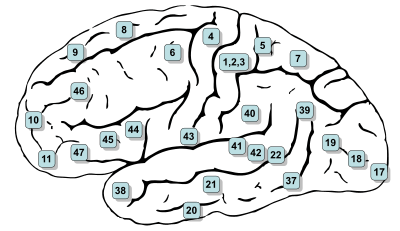 Frontal eye field is roughly located between regions #4, #6, and #8 | |
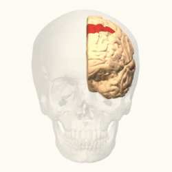
The frontal eye fields (FEF) are a region located in the frontal cortex, more specifically in Brodmann area 8 or BA8,[1] of the primate brain. In humans, it can be more accurately said to lie in a region around the intersection of the middle frontal gyrus with the precentral gyrus, consisting of a frontal and parietal portion.[2] The FEF is responsible for saccadic eye movements for the purpose of visual field perception and awareness, as well as for voluntary eye movement. The FEF communicates with extraocular muscles indirectly via the paramedian pontine reticular formation. Destruction to FEF causes deviation of the eyes to the ipsilateral side.
Function
The cortical area called frontal eye field (FEF) plays an important role in the control of visual attention and eye movements.[3] Electrical stimulation in the FEF elicits saccadic eye movements. The FEF have a topographic structure and represents saccade targets in retinotopic coordinates.[4]
The frontal eye field is reported to be activated during the initiation of eye movements, such as voluntary saccades[5] and pursuit eye movements.[6] There is also evidence that it plays a role in purely sensory processing and that it belongs to a “fast brain” system through a superior colliculus – medial dorsal nucleus – FEF ascending pathway.[7]
In humans, its earliest activations in regard to visual stimuli occur at 45 ms with activations related to changes in visual stimuli within 45–60 ms (these are comparable with response times in the primary visual cortex).[7] This fast brain pathway also provides auditory input at even shorter times starting at 24 ms and being affected by auditory characteristics at 30–60 ms.[7]
The FEF constitutes together with the supplementary eye fields (SEF), the intraparietal sulcus (IPS) and the superior colliculus (SC) one of the most important brain areas involved in the generation and control of eye movements, particularly in the direction contralateral to the frontal eye fields' location.
Clinical significance
Lesions
Unilateral stimulation of a FEF causes conjugate gaze contralateral to the stimulation. Therefore, a unilateral destructive lesion to a FEF causes conjugate gaze towards the lesion, while an irritative (stimulating) lesion of a FEF causes conjugate gaze away from the lesion.
See also
References
- ↑ "Frontal Eye Field--Scholarpedia".
- ↑ Vernet, M.; Quentin, R.; Chanes, L.; Mitsumasu, A.; Valero-Cabré, A. (2014). "Frontal eye field, where art thou? Anatomy, function, and non-invasive manipulation of frontal regions involved in eye movements and associated cognitive operations". Frontiers of Integrated Neuroscience. 8 (66). doi:10.3389/fnint.2014.00088. PMID 4141567.
- ↑ Schall, J. D. (2004). "On the role of frontal eye field in guiding attention and saccades". Vision Research. 44 (12): 1453–67. doi:10.1016/j.visres.2003.10.025. PMID 15066404.
- ↑ Bruce, C. J.; Goldberg, M. E.; Bushnell, M. C.; Stanton, G. B. (1985). "Primate frontal eye fields. II. Physiological and anatomical correlates of electrically evoked eye movements". Journal of Neurophysiology. 54 (3): 714–34. PMID 4045546.
- ↑ "Medical Neurosciences".
- ↑ Mustari MJ, Ono S, Das VE (May 2009). "Signal processing and distribution in cortical-brainstem pathways for smooth pursuit eye movements". Ann. N. Y. Acad. Sci. 1164: 147–54. doi:10.1111/j.1749-6632.2009.03859.x. PMC 3057571
 . PMID 19645893.
 - 1 2 3 Kirchner, H; Barbeau, E. J.; Thorpe, S. J.; Régis, J; Liégeois-Chauvel, C (2009). "Ultra-rapid sensory responses in the human frontal eye field region". Journal of Neuroscience. 29 (23): 7599–606. doi:10.1523/JNEUROSCI.1233-09.2009. PMID 19515928.
External links
| Wikimedia Commons has media related to Frontal eye field. |
| Related | ||
|---|---|---|