Feline leukemia virus
| Feline leukemia virus | |
|---|---|
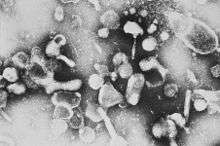 | |
| Electron micrograph of Feline leukemia virus | |
| Virus classification | |
| Group: | Group VI (ssRNA-RT) |
| Order: | Unassigned |
| Family: | Retroviridae |
| Subfamily: | Orthoretrovirinae |
| Genus: | Gammaretrovirus |
| Species: | Feline leukemia virus |
Feline leukemia virus (FeLV) is a retrovirus that infects cats. FeLV can be transmitted from infected cats when the transfer of saliva or nasal secretions is involved. If not defeated by the animal’s immune system, the virus can cause diseases which can be lethal. One disease caused by this virus is a form of cancer of the blood cells called Lymphoma.
Signs and symptoms
The signs and symptoms of infection with feline leukemia virus are quite varied and include loss of appetite, poor coat condition, anisocoria (uneven pupils), infections of the skin, bladder and respiratory tract, oral disease, seizures, lymphadenopathy (swollen lymph nodes), skin lesions, fatigue, fever, weight loss, stomatitis, gingivitis, litter box avoidance, pancytopenia, recurring bacterial and viral illnesses, anemia, diarrhea and jaundice.
Asymptomatic carriers will show no signs of disease, often for many years.
Progression
The disease has a wide range of effects. The cat can fight off the infection and become totally immune, can become a healthy carrier that never gets sick itself but can infect other cats, or a mid-level case in which the cat has a compromised immune system. Nevertheless, the development of lymphomas is considered the final stage of the disease. Although it is thought that virus protein has to be present to induce lymphomas in cats, newer evidence shows that a high percentage of FeLV-Antigen negative lymphomas contain FeLV-DNA, indicating a "hit-and-run" mechanism of virus-induced tumor development.[1]
Once the virus has entered the cat, there are six stages to a FeLV infection:
- Stage One: The virus enters the cat, usually through the pharynx where it infects the epithelial cells and infects the tonsilar B-lymphocytes and macrophages. These white blood cells then filter down to the lymph nodes and begin to replicate.
- Stage Two: The virus enters the blood stream and begins to distribute throughout the body.
- Stage Three: The lymphoid system (which produces antibodies to attack infected and cancerous cells) becomes infected, with further distribution throughout the body.
- Stage Four: The main point in the infection- where the virus can take over the body's immune system and cause viremia. During this stage the hemolymphatic system and intestines become infected.
If the cat's immune system does not fight off the virus, then it progresses to:
- Stage Five: The bone marrow becomes infected. At this point, the virus will stay with the cat for the rest of its life. In this phase, the virus replicates and is released four to seven days later in infected neutrophils, and sometimes lymphocytes, monocytes, and eosinophils (all white blood cells formed in the bone marrow).
- Stage Six: The cat's body is overwhelmed by infection and mucosal and glandular epithelial cells (tissue that forms a thin protective layer on exposed bodily surfaces and forms the lining of internal cavities, ducts, and organs) become infected. The virus replicates in epithelial tissues including salivary glands, oropharynx, stomach, esophagus, intestines, trachea, nasopharynx, renal tubules, bladder, pancreas, alveolar ducts, and sebaceous ducts from the muzzle.
Transmission
Cats infected with FeLV can serve as sources of infection. Transmission is related to the subgroup (see below). Cats can possibly pass the virus between themselves through saliva and close contact, by biting another cat, through a litter box or food dish used by an infected cat.
FeLV causes immunosuppression in domestic cats, and there is also evidence for existence of the virus in larger wild cat populations also (e.g. lynx, cheetahs, and lions). Overwhelming epidemiologic evidence suggests FeLV is not transmissible to either humans or dogs. This statement is based on the fact that approximately one pet dog in five lives with a cat, and many domestic cats live with humans (some 60 million pet cats in the United States). It is species-specific, and does not infect other animals, such as dogs (in fact, there is apparently no canine version of this disease at all).
Approximately 0.5% of pet cats are persistently infected with FeLV, but many more pet cats (>35%) have specific IgG antibodies which indicate prior exposure and subsequent development of immunity instead of infection. Transmission of FeLV is mainly via saliva during aggressive behaviors (fighting/biting) or exchange of bodily fluids (sexual contact/mating behavior). Friendly behaviors, such as sharing feeding bowls, water, bedding, or mutual grooming are believed to provide low risk of transmission.
Kittens can be born with it, having contracted it from their mother while in utero.
Infection is far higher in city cats, stray or owned, than in rural cats: this is entirely due to the amount of contact the cats have with each other.
Four subgroups of FeLV exist: A, B, C, and T, but only subgroup A is transmissible between cats. The other subgroups arise de novo and as results of recombination with an endogenous feline DNA sequence. Hence, there is very good evidence this virus is quite ancient, and may well have evolved more than one time over the last 10,000,000 years.
Subgroups are defined on the basis of viral interference and in vitro host range. The differences are due to polymorphism in the envelope glycoprotein gp70 with the highest level of divergence lying in the region of gp70 which is thought to interact with the cellular receptor. In an infected cell, gp70 is thought to block viral receptors, so preventing further infection by the same subgroup.
Diagnosis and prognosis
Cats diagnosed as persistently infected by ELISA testing may die within a few months or may remain asymptomatic for longer. The fatal diseases are leukemias, lymphomas, and non-regenerative anemias. Although there is no known cure for the virus infection, in 2006 the United States Department of Agriculture approved Lymphocyte T-Cell Immunomodulator as a treatment aid for FeLV (see Treatment).
Prevention
Vaccines for FeLV are available (ATCvet code QI06AA01 (WHO) and various combination vaccines), though no currently available vaccine offers 100% protection from the virus.[2] Serious side effects have also been reported as a result of FeLV vaccination; in particular, a small percentage of cats who received FeLV vaccines subsequently developed vaccine-associated sarcomas, an aggressive tumour, at the injection site.[3] The development of sarcomas with the use of the old FeLV and other vaccines may be due to the inflammation caused by aluminium adjuvants in the vaccines.[4]
Merial produces a recombinant vaccine consisting of canarypox virus carrying FeLV gag and env genes (sold as PUREVAX FeLV in the USA and Eurifel FeLV in Europe). This is thought to be safer than the old vaccine as it does not require an adjuvant to be effective. Although this is a live virus, it originates from a bird host and so does not replicate in mammals.[5]
Since the virus is very weak and dies within two hours in a dry environment, the incidence of transmission will drop considerably if the litterbox is kept from remaining damp between uses. One method is to clean all damp litter out of a standard box after each use; however, this is sometimes not practical.
Another option is a specialized three-part litterbox that uses either a ground corncob or safflower seed litter in a slotted top unit, which allows the liquid to drain into a reservoir that is emptied regularly. The litter material air-dries quickly, thus killing the virus quickly.
This litterbox was originally designed for diabetic cats, to allow regular testing of the sugar level in the cat's system. By coincidence, the box also helps prevent FeLV infection between household cats .
Viral structure
Feline Leukemia Virus (FeLV) is an RNA virus in the subfamily Oncovirinae belong to family Retroviridae was first described by W. Jarrett (et al., Nature 202:566) at University of Glasgow, School Veterinary Medicine, in 1964. The virus comprises 5' and 3' LTRs and three genes: Gag (structural), Pol (enzymes) and Env (envelope and transmembrane); the total genome is about 9,600 base pairs.
See the entry on retroviruses for more details on the life cycle of FeLV.
Treatment
Approved US treatment
In 2006, the United States Department of Agriculture issued a conditional license for a new treatment aid termed Lymphocyte T-Cell Immunomodulator (LTCI).[6] Lymphocyte T-Cell Immunomodulator is manufactured and distributed exclusively by T-Cyte Therapeutics, Inc.[7]
Lymphocyte T-Cell Immunomodulator is intended as an aid in the treatment of cats infected with feline leukemia virus (FeLV) and/or feline immunodeficiency virus (FIV), and the associated symptoms of lymphocytopenia, opportunistic infection, anemia, granulocytopenia, or thrombocytopenia. The absence of any observed adverse events in several animal species suggests that the product has a very low toxicity profile.
Lymphocyte T-Cell Immunomodulator is a potent regulator of CD-4 lymphocyte production and function.[8] It has been shown to increase lymphocyte numbers and Interleukin 2 production in animals.[9]
Lymphocyte T-Cell Immunomodulator is a single chain polypeptide. It is a strongly cationic glycoprotein, and is purified with cation exchange resin. Purification of protein from bovine-derived stromal cell supernatants produces a substantially homogeneous factor, free of extraneous materials. The bovine protein is homologous with other mammalian species and is a homogeneous 50 kDa glycoprotein with an isoelectric point of 6.5. The protein is prepared in a lyophilized 1 microgram dose. Reconstitution in sterile diluent produces a solution for subcutaneous injection.[7][10]
Approved European treatment
Interferon-ω (omega) is sold in Europe at least under the name Virbagen Omega and manufactured by Virbac. When used in treatment of cats infected with FeLV in non-terminal clinical stages (over the age of 9 weeks) there have been substantial improvements in mortality rates; in non-anemic cats, mortality rate of 50% was reduced by approximately 20% following treatment.
History
The name stems from the fact that the first disease associated with the virus was a form of leukemia. By the time it was discovered that the virus was not the same as leukemia, the misnomer had already found its way into the vocabulary of pet owners.
Comparison with feline immunodeficiency virus
FeLV and feline immunodeficiency virus (FIV) and are sometimes mistaken for one another though the viruses differ in many ways. Although they are both in the same retroviral family (orthoretrovirinea), they are classified in different genera (FeLV is a gamma-retrovirus and FIV is a lentivirus like HIV-1). Their shapes are quite different: FeLV is more circular while FIV is elongated. The two viruses are also quite different genetically, and their protein coats differ in size and composition. Although many of the diseases caused by FeLV and FIV are similar, the specific ways in which they are caused also differs. Also, while the feline leukemia virus may cause symptomatic illness in an infected cat, an FIV infected cat can remain completely asymptomatic its entire lifetime.
See also
References
- ↑ Weiss AT, Klopfleisch R, Gruber AD (2010). "Prevalence of feline leukaemia provirus DNA in feline lymphomas.". J Feline Med Surg. 12 (12): 929–35. doi:10.1016/j.jfms.2010.07.006. PMID 21036089.
- ↑ "Feline Leukemia Virus: A Cause of Immunodeficiency in Cats".
- ↑ "Feline Leukemia Virus Diseases".
- ↑ Richards J, Elston T, Ford R, Gaskell R, Hartmann K, Hurley K, Lappin M, Levy J, Rodan I, Scherk M, Schultz R, Sparkes A (2006). "The 2006 American Association of Feline Practitioners Feline Vaccine Advisory Panel report". J Am Vet Med Assoc. 229 (9): 1405–41. doi:10.2460/javma.229.9.1405. PMID 17078805.
- ↑ Poulet H, Brunet S, Boularand C, Guiot AL, Leroy V, Tartaglia J, Minke J, Audonnet JC, Desmettre P (2003). "Efficacy of a canarypox virus-vectored vaccine against feline leukaemia". The Veterinary Record. 153 (5): 141–5. doi:10.1136/vr.153.5.141. PMID 12934796.
- ↑ "LTCI Product Information". T-Cyte Therapeutics, Inc. Retrieved 28 July 2012.
- 1 2 "T-Cyte Therapeutics, Inc.". T-Cyte Therapeutics, Inc. Retrieved 28 July 2012.
- ↑ Beardsley, et al. "Induction of T-Cell Maturation by a Cloned Line of Thymic Epithelium (TEPI) Immunology 80: pp. 6005-6009, (Oct. 1983).
- ↑ Beardsley, Terry R. Patent # 7,196,060; Method to enhance hematopoiesis. Method to enhance hematopoiesis - Google Patents at www.google.com
- ↑ Beardsley, Terry R. Patent # 5,616,554; Immune-enhancing agent for therapeutic use in immunocompromised hosts. Immune-enhancing agent for ... - Google Patents at www.google.com
External links
- Lymphocyte T-Cell Immunomodulator (LTCI)
- Feline Leukemia Virus (FeLV) from Veterinary Partner
- Signs And Symptoms Of Feline Leukemia
- Feline leukemia Treatments and Support
- http://cyberleninka.ru/article/n/rentgenologicheskiy-metod-v-diagnostike-retrovirusnyh-infektsiy-koshek
- "Feline leukemia virus". NCBI Taxonomy Browser. 11768.