Fascia of Scarpa
| Fascia of Scarpa | |
|---|---|
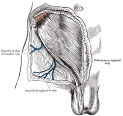 The subcutaneous inguinal ring. (Superficial fascia visible at top.) | |
| Details | |
| Identifiers | |
| Latin | membranous layer telae subcutaneae abdominis |
| TA | A04.5.02.022 |
The fascia of Scarpa is the deep membranous layer (stratum membranosum), of the superficial fascia of the abdomen. It is a layer of the anterior abdominal wall. It is found deep to the Camper Fascia and superficial to the external oblique muscle.
Structure
It is thinner and more membranous in character than the superficial fascia of Camper, and contains a considerable quantity of orange elastic fibers.
It is loosely connected by areolar tissue to the aponeurosis of the external oblique muscle, but in the midline it is more intimately adherent to the linea alba and to the pubic symphysis, and is prolonged on to the dorsum of the penis, forming the fundiform ligament; above, it is continuous with the superficial fascia over the rest of the trunk; below and laterally, it blends with the fascia lata of the thigh a little below the inguinal ligament; medially and below, it is continued over the penis and spermatic cord to the scrotum, where it helps to form the dartos.
From the scrotum it may be traced backward into continuity with the deep layer of the superficial fascia of the perineum (superficial perineal fascia or fascia of Colles).
In the female, it is continued into the labia majora and from there to the fascia of Colles.
History
It is named for Italian anatomist Antonio Scarpa.[1] Antonio Scarpa's description of the membranous superficial fascia is vague in his 1809 hernia monograph.[2] Life-size illustrations included by Scarpa do not identify the layer even though some show all the other anatomical layers of the abdominal wall in the inguinal region. A probable description of the fascia is in the text which discusses femoral (called crural) hernia in the male. Scarpa describes that 'below the skin' we find 'a layer of condensed substance forming the second covering of the hernia' which adheres to 'the aponeurosis of the fascia lata'. A little later he describes this layer as being membranous and he believes it has a role in containing this particular herniation. In 1810, Abraham Colles described detailed methods of dissection to expose membranous superficial fascia in the lower abdomen and the inguino-perineal region including the penis and scrotum. Colles clearly associated the subcutaneous limitation of urine extravasation from a ruptured urethra with the attachments of membranous superficial fascia to deeper structures.[3]
Clinical significance
Scarpa's belief that the fascia stops hernias from forming is not thought to be true today. Some anatomists suggest the membranous superficial fascia is the scaffold which attaches the skin to the deeper structures so that the skin does not sag with gravity but still stretches as the body flexes or changes shape with exercise.[3] The attachment of the fascia to deeper layers confines fluid which may have come from inside the body in certain diseases giving rise to clinical signs such as urethral disruption noticed by Colles and bruising in Cullen's sign or Grey Turner's sign.[3]
References
This article incorporates text in the public domain from the 20th edition of Gray's Anatomy (1918)
- ↑ synd/2925 at Who Named It?
- ↑ A. Scarpa. Sull' ernie: memorie anatomico-chirurgiche. Milano, d. reale Stamperia, 1809; 2nd edition, 1820.
- 1 2 3 Ullah, S. M.; Grant, R. C.; Johnson, M; McAlister, V. C. (2013). "Scarpa's fascia and clinical signs: The role of the membranous superficial fascia in the eponymous clinical signs of retroperitoneal catastrophe". Annals of the Royal College of Surgeons of England. 95 (7): 519–22. doi:10.1308/003588413X13629960048514. PMID 24112501.
External links
- Anatomy photo:35:03-0103 at the SUNY Downstate Medical Center
- Anatomy figure: 35:03-03 at Human Anatomy Online, SUNY Downstate Medical Center
- Atlas image: abdo_wall57 at the University of Michigan Health System
- Anatomy image:7410 at the SUNY Downstate Medical Center
- Anatomy image:7042 at the SUNY Downstate Medical Center