Falciform ligament
| Falciform ligament | |
|---|---|
 The superior surface of the liver. ("Attachment of falciform ligament" is white band to the left.) | |
 1: Right lobe of liver 2: Left lobe of liver 3: Quadrate lobe of liver 4: Round ligament of liver 5: Falciform ligament 6: Caudate lobe of liver 7: Inferior vena cava 8: Common bile duct 9: Hepatic artery 10: Portal vein 11: Cystic duct 12: Hepatic duct 13: Gallbladder | |
| Details | |
| Identifiers | |
| Latin | ligamentum falciforme hepatis |
| TA | A10.1.02.303 |
| FMA | 15823 |
The falciform ligament is a ligament that attaches the liver to the anterior (ventral) body wall. It is a broad and thin antero-posterior peritoneal fold, falciform (Latin "sickle-shaped"), its base being directed downward and backward and its apex upward and backward. The falciform ligament droops down from the hilum of the liver.
Structure
It is situated in an antero-posterior plane but lies obliquely, so that one surface faces forward and is in contact with the peritoneum behind the right rectus and the diaphragm, while the other is directed backward and is in contact with the left lobe of the liver.
It is attached by its left margin to the under surface of the diaphragm and the posterior surface of the sheath of the right Rectus as low down as the umbilicus; by its right margin it extends from the notch on the anterior margin of the liver, as far back as the posterior surface.
It is composed of two layers of peritoneum closely united together.
Its base or free edge contains between its layers the round ligament and the paraumbilical veins.
Development
It is a remnant of the umbilical vein of the fetus and a derivative of the embryonic ventral mesentery.
Clinical significance
Becomes canalised if the individual is suffering from portal hypertension. Due to the increase in venous congestion, blood is pushed down from the liver towards the anterior abdominal wall and if blood pools here, will result in periumbilical bruising.
Additional images
-

The primitive mesentery of a six weeks’ human embryo, half schematic.
-
Horizontal disposition of the peritoneum in the upper part of the abdomen.
-

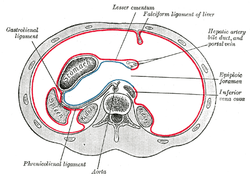
Diagram to show the lines along which the peritoneum leaves the wall of the abdomen to invest the viscera.
-

Falciform ligament.Superior surface of liver.
External links
- Anatomy photo:37:04-0100 at the SUNY Downstate Medical Center - "Abdominal Cavity: The Falciform Ligament of the Liver"
- Anatomy photo:38:12-0205 at the SUNY Downstate Medical Center - "The Visceral Surface of the Liver"
- Anatomy image:8373 at the SUNY Downstate Medical Center
- liver at The Anatomy Lesson by Wesley Norman (Georgetown University)
- Cross section image: pembody/body8a - Plastination Laboratory at the Medical University of Vienna
References
This article incorporates text in the public domain from the 20th edition of Gray's Anatomy (1918)