Extraglomerular mesangial cell
| Extraglomerular mesangial cell | |
|---|---|
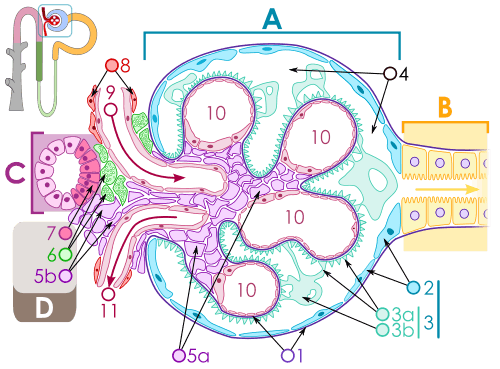 Diagram of renal corpuscle structure. Extraglomerular mesangial cell is #5b. A – Renal corpuscle B – Proximal tubule C – Distal convoluted tubule D – Juxtaglomerular apparatus 1. Basement membrane (Basal lamina) 2. Bowman's capsule – parietal layer 3. Bowman's capsule – visceral layer 3a. Pedicels (Foot processes from podocytes) 3b. Podocyte 4. Bowman's space (urinary space) 5a. Mesangium – Intraglomerular cell 5b. Mesangium – Extraglomerular cell 6. Granular cells (Juxtaglomerular cells) 7. Macula densa 8. Myocytes (smooth muscle) 9. Afferent arteriole 10. Glomerulus Capillaries 11. Efferent arteriole | |
Extraglomerular mesangial cells (also known as Lacis cells, Goormaghtigh cells, or Polkissen cells) are light-staining pericytes in the kidney found outside the glomerulus, near the vascular pole. They resemble smooth muscle cells and play a role in renal autoregulation of blood flow to the kidney and regulation of systemic blood pressure through the renin-angiotensin system. Extraglomerular mesangial cells are part of the juxtaglomerular apparatus, along with the macula densa cells of the distal convoluted tubule and the juxtaglomerular cells of the afferent arteriole.
The specific function of extraglomerular mesangial cells is not well understood, although it has been associated with the secretion of erythropoietin.[1] They are distinguished from intraglomerular mesangial cells, which are situated between the basement membrane and the epithelial cells within the glomerulus.