Echogenicity
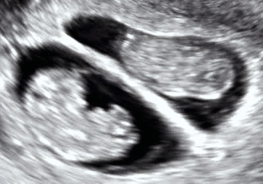
Echogenicity (misspelled sometimes as echogenecity) is the ability to bounce an echo, e.g. return the signal in ultrasound examinations. In other words, echogenicity is higher when the surface bouncing the sound echo reflects increased sound waves. Tissues that have higher echogenicity are called "hyperechogenic" and are usually represented with lighter colors on images in medical ultrasonography. In contrast, tissues with lower echogenicity are called "hypoechogenic" and are usually represented with darker colors. Areas that lack echogenicity are called "anechogenic" and are usually displayed as completely dark.
Microbubbles
Echogenicity can be increased by intravenously administering gas-filled microbubble contrast agent to the systemic circulation, with the procedure being called contrast-enhanced ultrasound. This is because microbubbles have a high degree of echogenicity. When gas bubbles are caught in an ultrasonic frequency field, they compress, oscillate, and reflect a characteristic echo- this generates the strong and unique sonogram in contrast-enhanced ultrasound. Gas cores can be composed of air, or heavy gases like perfluorocarbon, or nitrogen.[1] Heavy gases are less water-soluble so they are less likely to leak out from the microbubble to impair echogenicity (McCulloch et al., 2000). Therefore, microbubbles with heavy gas cores are likely to last longer in circulation.
Reasons for higher echogenicity
During ultrasound examinations, sometimes echogenicity is higher in certain parts of body. Fatty liver could cause increased echogenicity in the liver, especially if the liver transaminases are elevated.[2]
Women with polycystic ovary syndrome may also show an increase in stromal echogenicity
See also
References
- ↑ Lindner, JR (2004). "Microbubbles in medical imaging: current applications and future directions". Nature Reviews. Drug Discovery. 3 (6): 527–32. doi:10.1038/nrd1417.
- ↑ Mathiesen, UL; Franzén, LE. "Increased liver echogenicity at ultrasound examination reflects degree of steatosis but not of fibrosis in asymptomatic patients with mild/moderate abnormalities of liver transaminases". Scandinavian Journal of Gastroenterology. Department of Internal Medicine, County Hospital, Oskarshamn, Sweden. 34 (7): 516–22. doi:10.1016/s1590-8658(02)80111-6.