Echinococcus
| Echinococcus | |
|---|---|
 | |
| Scientific classification | |
| Kingdom: | Animalia |
| Phylum: | Platyhelminthes |
| Class: | Cestoda |
| Order: | Cyclophyllidea |
| Family: | Taeniidae |
| Genus: | Echinococcus |

The genus Echinococcus includes six parasite species of cyclophyllid tapeworms to date, of the family Taeniidae. Infection with Echinococcus results in hydatid disease, also known as echinococcosis.
Echinococcus is triploblastic – it has three layers – outermost ectoderm, middle mesoderm, and inner endoderm. An anus is absent, and it has no digestive system. Its body is covered by tegument and the worm is divided into a scolex, a short neck, and three to six proglottids. Its body shape is ribbon-like.
In humans, this causes a disease called echinococcosis. The three types of echinococcosis are cystic echinococcosis caused by E. granulosus, alveolar echinococcosis caused by E. multilocularis, and polycystic echinococcosis caused by E. vogeli or E. oligarthrus.[1] A worm's incubation period is usually long and can be up to 50 years. Cystic echinococcosis is mostly found in South and Central America, Africa, the Middle East, China, Italy, Spain, Greece, Russia, and the western United States (Arizona, New Mexico, and California).
Echinococcosis is a zoonosis. The definitive hosts are carnivorous predators – dogs, wolves, foxes, and lions. The adult tapeworm lives in their small intestines and delivers eggs to be excreted with the stool. The intermediate hosts are infected by ingesting eggs. Sheep, goats, cattle, camels, pigs, wild herbivores, and rodents are the usual intermediate hosts, but humans can also be infected. Humans are dead-end hosts, since their corpses are nowadays seldom eaten by carnivorous predators.
The egg hatches in the digestive system of the intermediate host, producing a planula larva. It penetrates the intestinal wall and is carried by bloodstream to liver, lung, brain, or another organ. It settles there and turns into a bladder-like structure called hydatid cyst. From the inner lining of its wall, protoscoleces (i.e. scoleces with invaginated tissue layers) bud and protrude into the fluid filling the cyst.
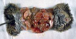
After the death of the normal intermediate host, its body can be eaten by carnivores suitable as definitive hosts. In their small intestines, protoscoleces turn inside out, attach, and give rise to adult tapeworms, completing the lifecycle. In humans, the cysts persist and grow for years. They are regularly found in the liver (and every possible organ: spleen, kidney, bone, brain, tongue and skin) and are asymptomatic until their growing size produces symptoms or are accidentally discovered. Disruption of the cysts (spontaneous or iatrogenic e.g. liver biopsy) can be life-threatening due to anaphylactic shock.
Cysts are detected with ultrasound, X-ray computed tomography, or other imaging techniques. Antiechinococcus antibodies can be detected with serodiagnostic tests – indirect fluorescent antibody, complement fixation, ELISA, Western blot, and other methods.[2]
Taxonomy
A phylogenetic tree has been created for several species in this genus – Echinococcus oligarthrus, Echinococcus vogeli, Echinococcus multilocularis, Echinococcus shiquicus, Echinococcus equinus, Echinococcus ortleppi, and Echinococcus granulosus.[3] The first diverging species are the neotropical endemic species E. oligarthrus and E. vogeli. E. ortleppi and E. canadensis are sister species, as are E. multilocularis and E. shiquicus. E. canadensis is related to E. granulosus.
The origin of these parasites based on host-parasite co-evolution comparisons was North America or Asia, depending on whether the ancestral definitive hosts were canids or felids.
Echinococcus oligarthrus and Echinococcus vogeli are basal in this genus.[4] The genus is a sister to the genus Taenia from which it diverged more than 10 million years ago. The genus Echinococcus evolved in North America in canids and began to diversify 5.8 million years ago.
Prevention
There is no vaccine against Echinococcus multilocularis. However it is possible to prevent humans from the fox tapeworm by deworming the main hosts.[5]
References
- ↑ Thompson RCA, McManus DP. Aetiology: parasites and life cycles. In: Eckert J, Gemmell MA, Meslin F-X, Pawlowski ZS editor. WHO/OIE Manual on Echinococcosis in Humans and Animals: a Public Health Problem of Global Concern. Paris: Office Internationale des Epizooities; 2001;p. 1–19
- ↑ Wenbao Zhang, Hao Wen, Jun Li, Renyong Lin, and Donald P. McManus, Immunology and Immunodiagnosis of Cystic Echinococcosis: An Update. Clinical and Developmental Immunology, 2012, 10 pages doi:10.1155/2012/101895
- ↑ Nakao M, McManus DP, Schantz PM, Craig PS, Ito A (2007) A molecular phylogeny of the genus Echinococcus inferred from complete mitochondrial genomes. Parasitology 134(5):713-722
- ↑ Knapp J, Nakao M, Yanagida T, Okamoto M, Saarma U, Lavikainen A, Ito A (2011) Phylogenetic relationships within Echinococcus and Taenia tapeworms (Cestoda: Taeniidae): an inference from nuclear protein-coding genes. Mol Phylogenet Evol 61(3):628-638. doi: 10.1016/j.ympev.2011.07.022
- ↑ Grabs A (November 2016). "How To Exterminate The Fox Tapeworm In Your Area".