Dunaliella salina
| Dunaliella salina | |
|---|---|
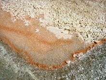 | |
| Orange-colored Dunaliella salina within sea salt | |
| Scientific classification | |
| Kingdom: | Plantae |
| Phylum: | Chlorophyta |
| Class: | Chlorophyceae |
| Order: | Chlamydomonadales |
| Family: | Dunaliellaceae |
| Genus: | Dunaliella |
| Species: | D. salina |
| Binomial name | |
| Dunaliella salina | |

Dunaliella salina is a type of halophile green micro-algae especially found in sea salt fields. Known for its antioxidant activity because of its ability to create large amount of carotenoids, it is used in cosmetics and dietary supplements. Few organisms can survive like D. salina does in such highly saline conditions as salt evaporation ponds. To survive, these organisms have high concentrations of β-carotene to protect against the intense light, and high concentrations of glycerol to provide protection against osmotic pressure. This offers an opportunity for commercial biological production of these substances.
History
Dunaliella salina was named by E.C. Teodoresco of Bucharest, Romania after its original discoverer, Michel Felix Dunal, who first scientifically reported sighting the organism in saltern evaporation ponds in Montpellier, France in 1838. He initially named the organism Haematoccocus salinus and Protococcus. The organism was fully described as a new, separate genus simultaneously by Teodoresco and Clara Hamburger of Heidelberg, Germany in 1905. Teodoresco was the first to publish his work, so he is generally given credit for this categorization.[1]
Morphology
Species in the Dunaliella genus are morphogically similar to Chlamydomonas reinhardtii with the main exception being that Dunaliella lack both a cell wall and a contractile vacuole. Dunaliella has two flagella of equal length and has a single cup-like chloroplast that often contains a central pyrenoid. The chloroplast can hold large amounts of β-carotene, which makes it appear orange-red. The β-carotene appears to protect the organism from long-term UV radiation that D. salina is exposed to in its typical environments. D. salina comes in various shapes and symmetries depending on the conditions in its current environment.[2]
Reproduction and lifecycle
D. salina can reproduce asexually through division of motile vegetative cells and sexually through the fusion of two equal gametes into a singular zygote. Though D. salina can survive in salinic environments, Martinez et al. determined that sexual activity of D. salina significantly decreases in higher salt concentrations (>10%) and is induced in lower salt concentrations.[3] Sexual reproduction begins when two D. salina’s flagella touch leading to gamete fusion. The D. salina zygote is extraordinarily hardy and can survive exposure to fresh water and to dryness. After germination, the zygotes release up to 32 haploid daughter cells.[4]
Commercial production of β-carotene
From a first pilot plant for D. salina cultivation for β-carotene production established in the USSR in 1966, the commercial cultivation of D. salina for the production of β-carotene throughout the world is now one of the success stories of halophile biotechnology.[5][6][7] Different technologies are used, from low-tech extensive cultivation in lagoons to intensive cultivation at high cell densities under carefully controlled conditions.[8]
Anti-oxidant & nutritional supplement
Due to the abundance of β-carotene, which is an anti-oxidant as well as a vitamin A precursor, D. salina is a popular pro-vitamin A food supplement and cosmetic additive.[9] Studies have demonstrated that D. salina exhibits strong protective effects on UVB radiation-induced corneal oxidative damage in mice, which seems to be caused by the increase of anti-oxidant enzyme activity and the inhibition of lipid peroxidation.Research performed at the Cancer Research Centre of Hawaii shows that D. salina contains a certain type of β-carotene called 9-cis-β-carotene, which is up to ten times stronger at preventing cancer than ordinary β-carotene.[10] Clinical studies have also shown that whole dried D. salina cells are an effective source of bioavailable carotenoids and 14 weeks of supplementation with D. salina can produce favourable shifts in heavy metals and elemental minerals in humans.[11]
Glycerol
D. salina lacks a rigid cell wall, which makes organism susceptible to osmotic pressure. Glycerol is used as a means by which to maintain both osmotic balance and enzymatic activity.[12] D. salina preserves a high concentration of glycerol by maintaining a cell membrane with low permeability to glycerol and synthesizing large quantities of glycerol from starch as a response to high extracellcular salt concentration, which is why it tends to thrive in highly salinic environments. Attempts have been made to exploit the high concentrations of glycerol accumulated by D. salina as the basis for the commercial production of this compound. Although technically the production of glycerol from D. salina was shown to be possible, economic feasibility is low and no biotechnological operation exists to exploit the alga for glycerol production.[13]
See also
| Wikimedia Commons has media related to Dunaliella salina. |
References
- ↑ Oren, Aharon (2005). "A hundred years of Dunaliella research: 1905-2005". Saline Systems. 1: 2. doi:10.1186/1746-1448-1-2. PMC 1224875
 . PMID 16176593.
 - ↑ BOROWITZKA, MICHAEL A. "THE MASS CULTURE OF DUNALIELLA SALINA". fao.org. Food and Agriculture Organization of the United Nations: Fisheries and Aquaculture Department. Retrieved 7 May 2016.
- ↑ Martinez, G.; Cifuentes, A.; Gonzalez, M.; Parra, O. (1995). "Effect of salinity on sexual activity of Dunaliella salina (Dunal) Teodoresco, strain CONC-006". Revista Chilena de Historia Natural.
- ↑ Lerche W. Untersuchungen über Entwicklung und Fortpflanzung in der Gattung Dunaliella. Arch f Protistenkd. 1937; 88:236–268.
- ↑ Ben-Amotz A. Glycerol, β-carotene and dry algal meal production by commercial cultivation of Dunaliella. In: Shelef G, Soeder CJ, editor. Algae Biomass. Amsterdam: Elsevier; 1980. pp. 603–610.
- ↑ Ben-Amotz A, Avron M. Accumulation of metabolites by halotolerant algae and its industrial potential. Ann Rev Microbiol. 1983;37:95–119. doi: 10.1146/annurev.mi.37.100183.000523.
- ↑ Borowitzka LJ, Borowitzka MA, Moulton TP. The mass culture of Dunaliella for fine chemicals: from laboratory to pilot plant. Hydrobiologia. 1984;116/117:115–121. doi: 10.1007/BF00027649.
- ↑ Ben-Amotz A, Avron M. The biotechnology of mass culturing Dunaliella for products of commercial interest. In: Cresswell RC, Rees TAV, Shah, N, editor. Algal and Cyanobacterial Biotechnology. Harlow: Longman Scientific and Technical Press; 1989. pp. 91–114.
- ↑ Mokady S, Abramovici A, Cogau U. The safety evaluation of Dunaliella bardawil as a potential food supplement. Food Chem Toxicol. 1989;27:221–6.
- ↑ Tsai, Chia-Fang, Fung-Jou Lu, and Yu-Wen Hsu. “Protective Effects of Dunaliella salina – a Carotenoids-Rich Alga – against Ultraviolet B-Induced Corneal Oxidative Damage in Mice.” Molecular Vision 18 (2012): 1540–1547. Print.
- ↑ Bobrov, Zac; Tracton, Ian; Taunton, Kathrine; Mathews, Dr. Megan. "Effectiveness of whole dried Dunaliella salina marine microalgae in the chelating and detoxification of toxic minerals and heavy metals." (PDF). interclinical.com.au. Retrieved 7 May 2016.
- ↑ Craigie JS, McLachlan J. Glycerol as a photosynthetic product in Dunaliella tertiolecta Butcher. Can J Bot. 1964;42:777–778.
- ↑ Chen BJ, Chi CH. Process development and evaluation for algal glycerol production. Biotechnol Bioengin. 1981;23:1267–1287. doi: 10.1002/bit.260230608.
Further reading
Borowitzka, M.J. & Siva, C.J. (2007). The taxonomy of the genus Dunaliella (Chlorophyta, Dunaliellales) with emphasis on the marine and halophilic species. Journal of Applied Phycology 19: 567-590.
Chen H., Lu Y. and Jiang J. “Comparative Analysis on the Key Enzymes of the Glycerol Cycle Metabolic Pathway in Dunaliella salina under Osmotic Stresses.” PLoS ONE, 2012, DOI: 10.1371/journal.pone.0037578
Massjuk, N.P. & Lilitska, G.G. (2011). Dunaliellales. In: Algae of Ukraine: diversity, nomenclature, taxonomy, ecology and geography. Volume 3: Chlorophyta. (Tsarenko, P.M., Wasser, S.P. & Nevo, E. Eds), pp. 152–157. Ruggell: A.R.A. Gantner Verlag K.-G..
Mixed Carotenoids. Rejuvenal healthy aging, n.d. Web. 22 Nov 2012.
Shariati M., Hadi M.R. “Microalgal Biotechnology and Bioenergy in Dunaliella” Biomedical Engineering, 2011. DOI: 10.5772/19046
Smith D., Lee R., Cushman J., Magnuson J., Tran D. and Polle J.” The Dunaliella salina organelle genomes: large sequences, inflated with intronic and intergenic DNA.” BMA Plant Biology, 2010. DOI: 10.1186/1471-2229-10-83
Tammam A., Fakhry E. and El-Sheekh M. “Effect of salt stress on antioxidant system and the metabolism of the reactive oxygen species in Dunaliella saline and Dunaliella tertiolecta.” African Journal of Biotechnology, 2011. DOI: 10.5897/AJB10.2392
Zhao, R., Cao, Y., Xu, H., Lv, L., Qiao, D. & Cao, Y. (2011). Analysis of expressed sequence tags from the green alga Dunaliella salina (Chlorophyta). Journal of Phycology 47(6): 1454-1460.
External links
- Dunaliella salina at the Encyclopedia of Life

- Guiry, M.D.; Guiry, G.M. (2008). "'Dunaliella salina'". AlgaeBase. World-wide electronic publication, National University of Ireland, Galway.
- MicrobeWiki reference on Dunaliella salina