Brain–computer interface
A brain–computer interface (BCI), sometimes called a mind-machine interface (MMI), direct neural interface (DNI), or brain–machine interface (BMI), is a direct communication pathway between an enhanced or wired brain and an external device. BCIs are often directed at researching, mapping, assisting, augmenting, or repairing human cognitive or sensory-motor functions.
Research on BCIs began in the 1970s at the University of California, Los Angeles (UCLA) under a grant from the National Science Foundation, followed by a contract from DARPA.[1][2] The papers published after this research also mark the first appearance of the expression brain–computer interface in scientific literature.
The field of BCI research and development has since focused primarily on neuroprosthetics applications that aim at restoring damaged hearing, sight and movement. Thanks to the remarkable cortical plasticity of the brain, signals from implanted prostheses can, after adaptation, be handled by the brain like natural sensor or effector channels.[3] Following years of animal experimentation, the first neuroprosthetic devices implanted in humans appeared in the mid-1990s.
History
The history of brain–computer interfaces (BCIs) starts with Hans Berger's discovery of the electrical activity of the human brain and the development of electroencephalography (EEG). In 1924 Berger was the first to record human brain activity by means of EEG. Berger was able to identify oscillatory activity, such as Berger's wave or the alpha wave (8–13 Hz), by analyzing EEG traces.
Berger's first recording device was very rudimentary. He inserted silver wires under the scalps of his patients. These were later replaced by silver foils attached to the patients' head by rubber bandages. Berger connected these sensors to a Lippmann capillary electrometer, with disappointing results. However, more sophisticated measuring devices, such as the Siemens double-coil recording galvanometer, which displayed electric voltages as small as one ten thousandth of a volt, led to success.
Berger analyzed the interrelation of alternations in his EEG wave diagrams with brain diseases. EEGs permitted completely new possibilities for the research of human brain activities.
Jacques Vidal coined the term "BCI" and produced the first peer-reviewed publications on this topic.[1][2] Vidal is widely recognized as the inventor of BCIs in the BCI community, as reflected in numerous peer-reviewed articles reviewing and discussing the field (e.g.,[4][5][6]).
Vidal's first BCI relied on visual evoked potentials to allow users to control cursor direction, and visual evoked potentials are still widely used in BCIs (Allison et al., 2010, 2012; Bin et al., 2011; Guger et al., 2012; Kaufmann et al., 2012; Jin et al., 2014; Kapeller et al., 2015).
After his early contributions, Vidal was not active in BCI research, nor BCI events such as conferences, for many years. In 2011, however, he gave a lecture in Graz, Austria, supported by the Future BNCI project, presenting the first BCI, which earned a standing ovation. Vidal was joined by his wife, Laryce Vidal, who previously worked with him at UCLA on his first BCI project. Prof. Vidal will also present a lecture on his early BCI work at the Sixth Annual BCI Meeting, scheduled for May–June 2016 at Asilomar, California.
Versus neuroprosthetics
Neuroprosthetics is an area of neuroscience concerned with neural prostheses, that is, using artificial devices to replace the function of impaired nervous systems and brain related problems, or of sensory organs. The most widely used neuroprosthetic device is the cochlear implant which, as of December 2010, had been implanted in approximately 220,000 people worldwide.[7] There are also several neuroprosthetic devices that aim to restore vision, including retinal implants.
The difference between BCIs and neuroprosthetics is mostly in how the terms are used: neuroprosthetics typically connect the nervous system to a device, whereas BCIs usually connect the brain (or nervous system) with a computer system. Practical neuroprosthetics can be linked to any part of the nervous system—for example, peripheral nerves—while the term "BCI" usually designates a narrower class of systems which interface with the central nervous system.
The terms are sometimes, however, used interchangeably. Neuroprosthetics and BCIs seek to achieve the same aims, such as restoring sight, hearing, movement, ability to communicate, and even cognitive function. Both use similar experimental methods and surgical techniques.
Animal BCI research
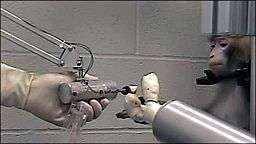
Several laboratories have managed to record signals from monkey and rat cerebral cortices to operate BCIs to produce movement. Monkeys have navigated computer cursors on screen and commanded robotic arms to perform simple tasks simply by thinking about the task and seeing the visual feedback, but without any motor output.[8] In May 2008 photographs that showed a monkey at the University of Pittsburgh Medical Center operating a robotic arm by thinking were published in a number of well-known science journals and magazines.[9] Other research on cats has decoded their neural visual signals.
Early work
In 1969 the operant conditioning studies of Fetz and colleagues, at the Regional Primate Research Center and Department of Physiology and Biophysics, University of Washington School of Medicine in Seattle, showed for the first time that monkeys could learn to control the deflection of a biofeedback meter arm with neural activity.[10] Similar work in the 1970s established that monkeys could quickly learn to voluntarily control the firing rates of individual and multiple neurons in the primary motor cortex if they were rewarded for generating appropriate patterns of neural activity.[11]
Studies that developed algorithms to reconstruct movements from motor cortex neurons, which control movement, date back to the 1970s. In the 1980s, Apostolos Georgopoulos at Johns Hopkins University found a mathematical relationship between the electrical responses of single motor cortex neurons in rhesus macaque monkeys and the direction in which they moved their arms (based on a cosine function). He also found that dispersed groups of neurons, in different areas of the monkey's brains, collectively controlled motor commands, but was able to record the firings of neurons in only one area at a time, because of the technical limitations imposed by his equipment.[12]
There has been rapid development in BCIs since the mid-1990s.[13] Several groups have been able to capture complex brain motor cortex signals by recording from neural ensembles (groups of neurons) and using these to control external devices.
Prominent research successes
Kennedy and Yang Dan
Phillip Kennedy (who later founded Neural Signals in 1987) and colleagues built the first intracortical brain–computer interface by implanting neurotrophic-cone electrodes into monkeys.

In 1999, researchers led by Yang Dan at the University of California, Berkeley decoded neuronal firings to reproduce images seen by cats. The team used an array of electrodes embedded in the thalamus (which integrates all of the brain’s sensory input) of sharp-eyed cats. Researchers targeted 177 brain cells in the thalamus lateral geniculate nucleus area, which decodes signals from the retina. The cats were shown eight short movies, and their neuron firings were recorded. Using mathematical filters, the researchers decoded the signals to generate movies of what the cats saw and were able to reconstruct recognizable scenes and moving objects.[14] Similar results in humans have since been achieved by researchers in Japan (see below).
Nicolelis
Miguel Nicolelis, a professor at Duke University, in Durham, North Carolina, has been a prominent proponent of using multiple electrodes spread over a greater area of the brain to obtain neuronal signals to drive a BCI.
After conducting initial studies in rats during the 1990s, Nicolelis and his colleagues developed BCIs that decoded brain activity in owl monkeys and used the devices to reproduce monkey movements in robotic arms. Monkeys have advanced reaching and grasping abilities and good hand manipulation skills, making them ideal test subjects for this kind of work.
By 2000 the group succeeded in building a BCI that reproduced owl monkey movements while the monkey operated a joystick or reached for food.[15] The BCI operated in real time and could also control a separate robot remotely over Internet protocol. But the monkeys could not see the arm moving and did not receive any feedback, a so-called open-loop BCI.
.jpg)
Later experiments by Nicolelis using rhesus monkeys succeeded in closing the feedback loop and reproduced monkey reaching and grasping movements in a robot arm. With their deeply cleft and furrowed brains, rhesus monkeys are considered to be better models for human neurophysiology than owl monkeys. The monkeys were trained to reach and grasp objects on a computer screen by manipulating a joystick while corresponding movements by a robot arm were hidden.[16][17] The monkeys were later shown the robot directly and learned to control it by viewing its movements. The BCI used velocity predictions to control reaching movements and simultaneously predicted handgripping force. In 2011 O'Doherty and colleagues showed a BCI with sensory feedback with rhesus monkeys. The monkey was brain controlling the position of an avatar arm while receiving sensory feedback through direct intracortical stimulation (ICMS) in the arm representation area of the sensory cortex.[18]
Donoghue, Schwartz and Andersen
Other laboratories which have developed BCIs and algorithms that decode neuron signals include those run by John Donoghue at Brown University, Andrew Schwartz at the University of Pittsburgh and Richard Andersen at Caltech. These researchers have been able to produce working BCIs, even using recorded signals from far fewer neurons than did Nicolelis (15–30 neurons versus 50–200 neurons).
Donoghue's group reported training rhesus monkeys to use a BCI to track visual targets on a computer screen (closed-loop BCI) with or without assistance of a joystick.[19] Schwartz's group created a BCI for three-dimensional tracking in virtual reality and also reproduced BCI control in a robotic arm.[20] The same group also created headlines when they demonstrated that a monkey could feed itself pieces of fruit and marshmallows using a robotic arm controlled by the animal's own brain signals.[21][22][23]
Andersen's group used recordings of premovement activity from the posterior parietal cortex in their BCI, including signals created when experimental animals anticipated receiving a reward.[24]
Other research
In addition to predicting kinematic and kinetic parameters of limb movements, BCIs that predict electromyographic or electrical activity of the muscles of primates are being developed.[25] Such BCIs could be used to restore mobility in paralyzed limbs by electrically stimulating muscles.
Miguel Nicolelis and colleagues demonstrated that the activity of large neural ensembles can predict arm position. This work made possible creation of BCIs that read arm movement intentions and translate them into movements of artificial actuators. Carmena and colleagues[16] programmed the neural coding in a BCI that allowed a monkey to control reaching and grasping movements by a robotic arm. Lebedev and colleagues[17] argued that brain networks reorganize to create a new representation of the robotic appendage in addition to the representation of the animal's own limbs.
The biggest impediment to BCI technology at present is the lack of a sensor modality that provides safe, accurate and robust access to brain signals. It is conceivable or even likely, however, that such a sensor will be developed within the next twenty years. The use of such a sensor should greatly expand the range of communication functions that can be provided using a BCI.
Development and implementation of a BCI system is complex and time consuming. In response to this problem, Gerwin Schalk has been developing a general-purpose system for BCI research, called BCI2000. BCI2000 has been in development since 2000 in a project led by the Brain–Computer Interface R&D Program at the Wadsworth Center of the New York State Department of Health in Albany, New York, United States.
A new 'wireless' approach uses light-gated ion channels such as Channelrhodopsin to control the activity of genetically defined subsets of neurons in vivo. In the context of a simple learning task, illumination of transfected cells in the somatosensory cortex influenced the decision making process of freely moving mice.[26]
The use of BMIs has also led to a deeper understanding of neural networks and the central nervous system. Research has shown that despite the inclination of neuroscientists to believe that neurons have the most effect when working together, single neurons can be conditioned through the use of BMIs to fire at a pattern that allows primates to control motor outputs. The use of BMIs has led to development of the single neuron insufficiency principle which states that even with a well tuned firing rate single neurons can only carry a narrow amount of information and therefore the highest level of accuracy is achieved by recording firings of the collective ensemble. Other principles discovered with the use of BMIs include the neuronal multitasking principle, the neuronal mass principle, the neural degeneracy principle, and the plasticity principle.[27]
BCIs are also proposed to be applied by users without disabilities. A user-centered categorization of BCI approaches by Thorsten O. Zander and Christian Kothe introduces the term passive BCI.[28] Next to active and reactive BCI that are used for directed control, passive BCIs allow for assessing and interpreting changes in the user state during Human-Computer Interaction (HCI). In a secondary, implicit control loop the computer system adapts to its user improving its usability in general.
The BCI Award
The Annual BCI Research Award, endowed with 3,000 USD, is awarded in recognition of outstanding and innovative research in the field of Brain-Computer Interfaces. Each year, a renowned research laboratory is asked to judge the submitted projects and to award the prize. The jury consists of world-leading BCI experts recruited by the awarding laboratory. Following list consists the winners of the BCI Award:[29]
- Motor imagery-based Brain-Computer Interface robotic rehabilitation for stroke.
- 2011: Moritz Grosse-Wentrup and Bernhard Schölkopf, (Max Planck Institute for Intelligent Systems, Germany)
- What are the neuro-physiological causes of performance variations in brain-computer interfacing?
- 2012: Surjo R. Soekadar and Niels Birbaumer, (Applied Neurotechnology Lab, University Hospital Tübingen and Institute of Medical Psychology and Behavioral Neurobiology, Eberhard Karls University, Tübingen, Germany)
- Improving Efficacy of Ipsilesional Brain-Computer Interface Training in Neurorehabilitation of Chronic Stroke
- 2013: M. C. Dadarlata,b, J. E. O’Dohertya, P. N. Sabesa,b (aDepartment of Physiology, Center for Integrative Neuroscience, San Francisco, CA, US, bUC Berkeley-UCSF Bioengineering Graduate Program, University of California, San Francisco, CA, US)
- A learning-based approach to artificial sensory feedback: intracortical microstimulation replaces and augments vision
- 2014: Katsuhiko Hamada, Hiromu Mori, Hiroyuki Shinoda, Tomasz M. Rutkowski, (The University of Tokyo, JP, Life Science Center of TARA, University of Tsukuba, JP, RIKEN Brain Science Institute, JP)
- Airborne Ultrasonic Tactile Display BCI
- 2015: Guy Hotson, David P McMullen, Matthew S. Fifer, Matthew S. Johannes, Kapil D. Katyal, Matthew P. Para, Robert Armiger, William S. Anderson, Nitish V. Thakor, Brock A. Wester, Nathan E. Crone (Johns Hopkins University, USA)
- Individual Finger Control of the Modular Prosthetic Limb using High-Density Electrocorticography in a Human Subject
- 2016: Gaurav Sharma, Nick Annetta, Dave Friedenberg, Marcie Bockbrader, Ammar Shaikhouni, W. Mysiw, Chad Bouton, Ali Rezai (Battelle Memorial Institute, The Ohio State University, USA)
- An Implanted BCI for Real-Time Cortical Control of Functional Wrist and Finger Movements in a Human with Quadriplegia
Human BCI research
Invasive BCIs
Vision
Invasive BCI research has targeted repairing damaged sight and providing new functionality for people with paralysis. Invasive BCIs are implanted directly into the grey matter of the brain during neurosurgery. Because they lie in the grey matter, invasive devices produce the highest quality signals of BCI devices but are prone to scar-tissue build-up, causing the signal to become weaker, or even non-existent, as the body reacts to a foreign object in the brain.[30]
In vision science, direct brain implants have been used to treat non-congenital (acquired) blindness. One of the first scientists to produce a working brain interface to restore sight was private researcher William Dobelle.
Dobelle's first prototype was implanted into "Jerry", a man blinded in adulthood, in 1978. A single-array BCI containing 68 electrodes was implanted onto Jerry’s visual cortex and succeeded in producing phosphenes, the sensation of seeing light. The system included cameras mounted on glasses to send signals to the implant. Initially, the implant allowed Jerry to see shades of grey in a limited field of vision at a low frame-rate. This also required him to be hooked up to a mainframe computer, but shrinking electronics and faster computers made his artificial eye more portable and now enable him to perform simple tasks unassisted.[31]
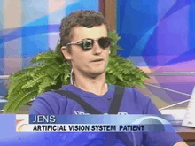
In 2002, Jens Naumann, also blinded in adulthood, became the first in a series of 16 paying patients to receive Dobelle’s second generation implant, marking one of the earliest commercial uses of BCIs. The second generation device used a more sophisticated implant enabling better mapping of phosphenes into coherent vision. Phosphenes are spread out across the visual field in what researchers call "the starry-night effect". Immediately after his implant, Jens was able to use his imperfectly restored vision to drive an automobile slowly around the parking area of the research institute.[32] Unfortunately, Dobelle died in 2004[33] before his processes and developments were documented. Subsequently, when Mr. Naumann and the other patients in the program began having problems with their vision, there was no relief and they eventually lost their "sight" again. Naumann wrote about his experience with Dobelle's work in Search for Paradise: A Patient's Account of the Artificial Vision Experiment[32] and has returned to his farm in Southeast Ontario, Canada, to resume his normal activities.[34]
Movement
BCIs focusing on motor neuroprosthetics aim to either restore movement in individuals with paralysis or provide devices to assist them, such as interfaces with computers or robot arms.
Researchers at Emory University in Atlanta, led by Philip Kennedy and Roy Bakay, were first to install a brain implant in a human that produced signals of high enough quality to simulate movement. Their patient, Johnny Ray (1944–2002), suffered from ‘locked-in syndrome’ after suffering a brain-stem stroke in 1997. Ray’s implant was installed in 1998 and he lived long enough to start working with the implant, eventually learning to control a computer cursor; he died in 2002 of a brain aneurysm.[35]
Tetraplegic Matt Nagle became the first person to control an artificial hand using a BCI in 2005 as part of the first nine-month human trial of Cyberkinetics’s BrainGate chip-implant. Implanted in Nagle’s right precentral gyrus (area of the motor cortex for arm movement), the 96-electrode BrainGate implant allowed Nagle to control a robotic arm by thinking about moving his hand as well as a computer cursor, lights and TV.[36] One year later, professor Jonathan Wolpaw received the prize of the Altran Foundation for Innovation to develop a Brain Computer Interface with electrodes located on the surface of the skull, instead of directly in the brain.
More recently, research teams led by the Braingate group at Brown University[37] and a group led by University of Pittsburgh Medical Center,[38] both in collaborations with the United States Department of Veterans Affairs, have demonstrated further success in direct control of robotic prosthetic limbs with many degrees of freedom using direct connections to arrays of neurons in the motor cortex of patients with tetraplegia.
Partially invasive BCIs
Partially invasive BCI devices are implanted inside the skull but rest outside the brain rather than within the grey matter. They produce better resolution signals than non-invasive BCIs where the bone tissue of the cranium deflects and deforms signals and have a lower risk of forming scar-tissue in the brain than fully invasive BCIs. There has been preclinical demonstration of intracortical BCIs from the stroke perilesional cortex.[39]
Electrocorticography (ECoG) measures the electrical activity of the brain taken from beneath the skull in a similar way to non-invasive electroencephalography (see below), but the electrodes are embedded in a thin plastic pad that is placed above the cortex, beneath the dura mater.[40] ECoG technologies were first trialled in humans in 2004 by Eric Leuthardt and Daniel Moran from Washington University in St Louis. In a later trial, the researchers enabled a teenage boy to play Space Invaders using his ECoG implant.[41] This research indicates that control is rapid, requires minimal training, and may be an ideal tradeoff with regards to signal fidelity and level of invasiveness.
(Note: these electrodes had not been implanted in the patient with the intention of developing a BCI. The patient had been suffering from severe epilepsy and the electrodes were temporarily implanted to help his physicians localize seizure foci; the BCI researchers simply took advantage of this.)[42]
Signals can be either subdural or epidural, but are not taken from within the brain parenchyma itself. It has not been studied extensively until recently due to the limited access of subjects. Currently, the only manner to acquire the signal for study is through the use of patients requiring invasive monitoring for localization and resection of an epileptogenic focus.
ECoG is a very promising intermediate BCI modality because it has higher spatial resolution, better signal-to-noise ratio, wider frequency range, and less training requirements than scalp-recorded EEG, and at the same time has lower technical difficulty, lower clinical risk, and probably superior long-term stability than intracortical single-neuron recording. This feature profile and recent evidence of the high level of control with minimal training requirements shows potential for real world application for people with motor disabilities.[43][44]
Light reactive imaging BCI devices are still in the realm of theory. These would involve implanting a laser inside the skull. The laser would be trained on a single neuron and the neuron's reflectance measured by a separate sensor. When the neuron fires, the laser light pattern and wavelengths it reflects would change slightly. This would allow researchers to monitor single neurons but require less contact with tissue and reduce the risk of scar-tissue build-up.
In 2014, a BCI study using near-infrared spectroscopy for "locked-in" patients with amyotrophic lateral sclerosis (ALS) was able to restore some basic ability of the patients to communicate with other people.[45]
Non-invasive BCIs
There have also been experiments in humans using non-invasive neuroimaging technologies as interfaces. The substantial majority of published BCI work involves noninvasive EEG-based BCIs. Noninvasive EEG-based technologies and interfaces have been used for a much broader variety of applications. Although EEG-based interfaces are easy to wear and do not require surgery, they have relatively poor spatial resolution and cannot effectively use higher-frequency signals because the skull dampens signals, dispersing and blurring the electromagnetic waves created by the neurons. EEG-based interfaces also require some time and effort prior to each usage session, whereas non-EEG-based ones, as well as invasive ones require no prior-usage training. Overall, the best BCI for each user depends on numerous factors.
Non-EEG-based Human-Computer Interface
Pupil-size oscillation
In a recent 2016 article, an entirely new communication device and non-EEG-based human-computer interface was developed, requiring no visual fixation or ability to move eyes at all, that is based on covert interest in (i.e. without fixing eyes on) chosen letter on a virtual keyboard with letters each having its own (background) circle that is micro-oscillating in brightness in different time transitions, where the letter selection is based on best fit between, on one hand, unintentional pupil-size oscillation pattern, and, on the other hand, the circle-in-background's brightness oscillation pattern. Accuracy is additionally improved by user's mental rehearsing the words 'bright' and 'dark' in synchrony with the brightness transitions of the circle/letter.[46]
Electroencephalography (EEG)-based brain-computer interfaces
Overview
Electroencephalography (EEG) is the most studied non-invasive interface, mainly due to its fine temporal resolution, ease of use, portability and low set-up cost. The technology is somewhat susceptible to noise however.
In the early days of BCI research, another substantial barrier to using EEG as a brain–computer interface was the extensive training required before users can work the technology. For example, in experiments beginning in the mid-1990s, Niels Birbaumer at the University of Tübingen in Germany trained severely paralysed people to self-regulate the slow cortical potentials in their EEG to such an extent that these signals could be used as a binary signal to control a computer cursor.[47] (Birbaumer had earlier trained epileptics to prevent impending fits by controlling this low voltage wave.) The experiment saw ten patients trained to move a computer cursor by controlling their brainwaves. The process was slow, requiring more than an hour for patients to write 100 characters with the cursor, while training often took many months. However, the slow cortical potential approach to BCIs has not been used in several years, since other approaches require little or no training, are faster and more accurate, and work for a greater proportion of users.
Another research parameter is the type of oscillatory activity that is measured. Birbaumer's later research with Jonathan Wolpaw at New York State University has focused on developing technology that would allow users to choose the brain signals they found easiest to operate a BCI, including mu and beta rhythms.
A further parameter is the method of feedback used and this is shown in studies of P300 signals. Patterns of P300 waves are generated involuntarily (stimulus-feedback) when people see something they recognize and may allow BCIs to decode categories of thoughts without training patients first. By contrast, the biofeedback methods described above require learning to control brainwaves so the resulting brain activity can be detected.
Lawrence Farwell and Emanuel Donchin developed an EEG-based brain–computer interface in the 1980s.[48] Their "mental prosthesis" used the P300 brainwave response to allow subjects, including one paralyzed Locked-In syndrome patient, to communicate words, letters and simple commands to a computer and thereby to speak through a speech synthesizer driven by the computer. A number of similar devices have been developed since then. In 2000, for example, research by Jessica Bayliss at the University of Rochester showed that volunteers wearing virtual reality helmets could control elements in a virtual world using their P300 EEG readings, including turning lights on and off and bringing a mock-up car to a stop.[49]
While an EEG based brain-computer interface has been pursued extensively by a number of research labs, recent advancements made by Bin He and his team at the University of Minnesota suggest the potential of an EEG based brain-computer interface to accomplish tasks close to invasive brain-computer interface. Using advanced functional neuroimaging including BOLD functional MRI and EEG source imaging, Bin He and co-workers identified the co-variation and co-localization of electrophysiological and hemodynamic signals induced by motor imagination.[50] Refined by a neuroimaging approach and by a training protocol, Bin He and co-workers demonstrated the ability of a non-invasive EEG based brain-computer interface to control the flight of a virtual helicopter in 3-dimensional space, based upon motor imagination.[51] In June 2013 it was announced that Bin He had developed the technique to enable a remote-control helicopter to be guided through an obstacle course.[52]
In addition to a brain-computer interface based on brain waves, as recorded from scalp EEG electrodes, Bin He and co-workers explored a virtual EEG signal-based brain-computer interface by first solving the EEG inverse problem and then used the resulting virtual EEG for brain-computer interface tasks. Well-controlled studies suggested the merits of such a source analysis based brain-computer interface.[53]
A 2014 study found that severely motor-impaired patients could communicate faster and more reliably with non-invasive EEG BCI, than with any muscle-based communication channel.[54]
Dry active electrode arrays
In the early 1990s Babak Taheri, at University of California, Davis demonstrated the first single and also multichannel dry active electrode arrays using micro-machining. The single channel dry EEG electrode construction and results were published in 1994.[55] The arrayed electrode was also demonstrated to perform well compared to silver/silver chloride electrodes. The device consisted of four sites of sensors with integrated electronics to reduce noise by impedance matching. The advantages of such electrodes are: (1) no electrolyte used, (2) no skin preparation, (3) significantly reduced sensor size, and (4) compatibility with EEG monitoring systems. The active electrode array is an integrated system made of an array of capacitive sensors with local integrated circuitry housed in a package with batteries to power the circuitry. This level of integration was required to achieve the functional performance obtained by the electrode.
The electrode was tested on an electrical test bench and on human subjects in four modalities of EEG activity, namely: (1) spontaneous EEG, (2) sensory event-related potentials, (3) brain stem potentials, and (4) cognitive event-related potentials. The performance of the dry electrode compared favorably with that of the standard wet electrodes in terms of skin preparation, no gel requirements (dry), and higher signal-to-noise ratio.[56]
In 1999 researchers at Case Western Reserve University, in Cleveland, Ohio, led by Hunter Peckham, used 64-electrode EEG skullcap to return limited hand movements to quadriplegic Jim Jatich. As Jatich concentrated on simple but opposite concepts like up and down, his beta-rhythm EEG output was analysed using software to identify patterns in the noise. A basic pattern was identified and used to control a switch: Above average activity was set to on, below average off. As well as enabling Jatich to control a computer cursor the signals were also used to drive the nerve controllers embedded in his hands, restoring some movement.[57]
Prosthesis and environment control
Non-invasive BCIs have also been applied to enable brain-control of prosthetic upper and lower extremity devices in people with paralysis. For example, Gert Pfurtscheller of Graz University of Technology and colleagues demonstrated a BCI-controlled functional electrical stimulation system to restore upper extremity movements in a person with tetraplegia due to spinal cord injury.[58] Between 2012 and 2013, researchers at the University of California, Irvine demonstrated for the first time that it is possible to use BCI technology to restore brain-controlled walking after spinal cord injury. In their spinal cord injury research study, a person with paraplegia was able to operate a BCI-robotic gait orthosis to regain basic brain-controlled ambulation.[59][60] In 2009 Alex Blainey, an independent researcher based in the UK, successfully used the Emotiv EPOC to control a 5 axis robot arm.[61] He then went on to make several demonstration mind controlled wheelchairs and home automation that could be operated by people with limited or no motor control such as those with paraplegia and cerebral palsy.
Other research
Electronic neural networks have been deployed which shift the learning phase from the user to the computer. Experiments by scientists at the Fraunhofer Society in 2004 using neural networks led to noticeable improvements within 30 minutes of training.[62]
Experiments by Eduardo Miranda, at the University of Plymouth in the UK, has aimed to use EEG recordings of mental activity associated with music to allow the disabled to express themselves musically through an encephalophone.[63] Ramaswamy Palaniappan has pioneered the development of BCI for use in biometrics to identify/authenticate a person.[64] The method has also been suggested for use as PIN generation device (for example in ATM and internet banking transactions.[65] The group which is now at University of Wolverhampton has previously developed analogue cursor control using thoughts.[66]
Researchers at the University of Twente in the Netherlands have been conducting research on using BCIs for non-disabled individuals, proposing that BCIs could improve error handling, task performance, and user experience and that they could broaden the user spectrum.[67] They particularly focused on BCI games,[68] suggesting that BCI games could provide challenge, fantasy and sociality to game players and could, thus, improve player experience.[69]
The first BCI session with 100% accuracy (based on 80 right-hand and 80 left-hand movement imaginations) was recorded in 1998 by Christoph Guger. The BCI system used 27 electrodes overlaying the sensorimotor cortex, weighted the electrodes with Common Spatial Patterns, calculated the running variance and used a linear discriminant analysis.[70]
Research is ongoing into military use of BCIs and since the 1970s DARPA has been funding research on this topic.[1][2] The current focus of research is user-to-user communication through analysis of neural signals.[71] The project "Silent Talk" aims to detect and analyze the word-specific neural signals, using EEG, which occur before speech is vocalized, and to see if the patterns are generalizable.[72]
DIY and open source BCI
In 2001, The OpenEEG Project[73] was initiated by a group of DIY neuroscientists and engineers. The ModularEEG was the primary device created the OpenEEG community; it was a 6-channel signal capture board that cost between $200 and $400 to make at home. The OpenEEG Project marked a significant moment in the emergence of DIY brain-computer interfacing.
In 2010, the Frontier Nerds of NYU's ITP program published a thorough tutorial titled How To Hack Toy EEGs.[74] The tutorial, which stirred the minds of many budding DIY BCI enthusiasts, demonstrated how to create a single channel at-home EEG with an Arduino and a Mattel Mindflex at a very reasonable price. This tutorial amplified the DIY BCI movement.
In 2013, OpenBCI emerged from a DARPA solicitation and subsequent Kickstarter campaign. They created a high-quality, open-source 8-channel EEG acquisition board, known as the 32bit Board, that retailed for under $500. Two years later they created the first 3D-printed EEG Headset, known as the Ultracortex, as well as, a 4-channel EEG acquisition board, known as the Ganglion Board, that retailed for under $100.
In 2015, NeuroTechX was created with the mission of building an international network for neurotechnology. They bring hackers, researchers and enthusiasts all together in many different cities around the world. According to their rapid growth, the DIY neurotech / BCI community was already waiting for such initiative to see light.
MEG and MRI

Magnetoencephalography (MEG) and functional magnetic resonance imaging (fMRI) have both been used successfully as non-invasive BCIs.[75] In a widely reported experiment, fMRI allowed two users being scanned to play Pong in real-time by altering their haemodynamic response or brain blood flow through biofeedback techniques.[76]
fMRI measurements of haemodynamic responses in real time have also been used to control robot arms with a seven-second delay between thought and movement.[77]
In 2008 research developed in the Advanced Telecommunications Research (ATR) Computational Neuroscience Laboratories in Kyoto, Japan, allowed the scientists to reconstruct images directly from the brain and display them on a computer in black and white at a resolution of 10x10 pixels. The article announcing these achievements was the cover story of the journal Neuron of 10 December 2008.[78]
In 2011 researchers from UC Berkeley published[79] a study reporting second-by-second reconstruction of videos watched by the study's subjects, from fMRI data. This was achieved by creating a statistical model relating visual patterns in videos shown to the subjects, to the brain activity caused by watching the videos. This model was then used to look up the 100 one-second video segments, in a database of 18 million seconds of random YouTube videos, whose visual patterns most closely matched the brain activity recorded when subjects watched a new video. These 100 one-second video extracts were then combined into a mashed-up image that resembled the video being watched.[80][81][82]
Neurogaming
Currently, there is a new field of gaming called Neurogaming, which uses non-invasive BCI in order to improve gameplay so that users can interact with a console without the use of a traditional controller.[83] Some Neurogaming software use a player's brain waves, heart rate, expressions, pupil dilation, and even emotions to complete tasks or affect the mood of the game.[84] For example, game developers at Emotiv have created non-invasive BCI that will determine the mood of a player and adjust music or scenery accordingly. This new form of interaction between player and software will enable a player to have a more realistic gaming experience.[85] Because there will be less disconnect between a player and console, Neurogaming will allow individuals to utilize their "psychological state"[86] and have their reactions transfer to games in real-time.[85]
However, since Neurogaming is still in its first stages, not much is written about the new industry. The first NeuroGaming Conference was held in San Francisco on May 1–2, 2013.[87]
BCI control strategies in neurogaming
Motor imagery
Motor imagery involves the imagination of the movement of various body parts resulting in sensorimotor cortex activation,which modulates sensorimotor oscillations in the EEG. This can be detected by the BCI to infer a user’s intent. Motor imagery typically requires a number of sessions of training before acceptable control of the BCI is acquired. These training sessions may take a number of hours over several days before users can consistently employ the technique with acceptable levels of precision. Regardless of the duration of the training session, users are unable to master the control scheme. This results in very slow pace of the gameplay.[88] Advance machine learning methods were recently developed to compute a subject-specific model for detecting the performance of motor imagery. The top performing algorithm from BCI Competition IV (http://www.bbci.de/competition/iv/) dataset 2 for motor imagery is the Filter Bank Common Spatial Pattern, developed by Ang et al. from A*STAR, Singapore).[89]
Bio/neurofeedback for passive BCI designs
Biofeedback is used to monitor a subject’s mental relaxation. In some cases, biofeedback does not monitor electroencephalography (EEG), but instead bodily parameters such as electromyography(EMG), galvanic skin resistance (GSR), and heart rate variability (HRV).Many biofeedback systems are used to treat certain disorders such as attention deficit hyperactivity disorder (ADHD), sleep problems in children, teeth grinding, and chronic pain. EEG biofeedback systems typically monitor four different bands (theta: 4–7 Hz, alpha:8–12 Hz, SMR: 12–15 Hz, beta: 15–18 Hz) and challenge the subject to control them. Passive BCI[28] involves using BCI to enrich human–machine interaction with implicit information on the actual user’s state, for example, simulations to detect when users intend to push brakes during an emergency car stopping procedure. Game developers using passive BCIs need to acknowledge that through repetition of game levels the user’s cognitive state will change or adapt. Within the first play of a level, the user will react to things differently from during the second play: for example, the user will be less surprised at an event in the game if he/she is expecting it.[88]
Visual Evoked Potential (VEP)
A VEP is an electrical potential recorded after a subject is presented with a type of visual stimuli. There are several types of VEPs.
Steady-state visually evoked potentials (SSVEPs) use potentials generated by exciting the retina, using visual stimuli modulated at certain frequencies. SSVEP’s stimuli are often formed from alternating checkerboard patterns and at times simply use flashing images . The frequency of the phase reversal of the stimulus used can be clearly distinguished in the spectrum of an EEG; this makes detection of SSVEP stimuli relatively easy . SSVEP has proved to be successful within many BCI systems . This is due to several factors, the signal elicited is measurable in as large a population as the transient VEP and blink movement and electro cardiographic artefacts do not affect the frequencies monitored. In addition, the SSVEP signal is exceptionally robust; the topographic organization of the primary visual cortex is such that a broader area obtains afferents from the central or fovial region of the visual field .SSVEP does have several problems however. As SSVEPs use flashing stimuli to infer a user’s intent, the user must gaze at one of the flashing or iterating symbols in order to interact with the system. It is, therefore, likely that the symbols could become irritating and uncomfortable to use during longer play sessions, which can often last more than an hour which may not be an ideal gameplay.
Another type of VEP used with applications is the P300 potential. The P300 event-related potential is a positive peak in the EEG that occurs at roughly 300 ms after the appearance of a target stimulus (a stimulus for which the user is waiting or seeking) or oddball stimuli . The P300 amplitude decreases as the target stimuli and the ignored stimuli grow more similar.The P300 is thought to be related to a higher level attention process or an orienting response Using P300 as a control scheme has the advantage of the participant only having to attend limited training sessions. The first application to use the P300 model was the P300 matrix . Within this system, a subject would choose a letter from a grid of 6 by 6 letters and numbers. The rows and columns of the grid flashed sequentially and every time the selected “choice letter” was illuminated the user’s P300 was (potentially) elicited. However, the communication process, at approximately 17 characters per minute, was quite slow. The P300 is a BCI that offers a discrete selection rather than a continuous control mechanism. The advantage of P300 use within games is that the player does not have to teach himself/herself how to use a completely new control system and so only has to undertake short training instances, to learn the gameplay mechanics and basic use of the BCI paradigm.[88]
Synthetic telepathy/silent communication
In a $6.3 million Army initiative to invent devices for telepathic communication, Gerwin Schalk, underwritten in a $2.2 million grant, found that it is possible to use ECoG signals to discriminate the vowels and consonants embedded in spoken and in imagined words. The results shed light on the distinct mechanisms associated with production of vowels and consonants, and could provide the basis for brain-based communication using imagined speech.[44][90]
Research into synthetic telepathy using subvocalization is taking place at the University of California, Irvine under lead scientist Mike D'Zmura. The first such communication took place in the 1960s using EEG to create Morse code using brain alpha waves. Using EEG to communicate imagined speech is less accurate than the invasive method of placing an electrode between the skull and the brain.[91] On February 27, 2013 the group of Miguel Nicolelis at Duke University and IINN-ELS successfully connected the brains of two rats with electronic interfaces that allowed them to directly share information, in the first-ever direct brain-to-brain interface.[92][93][94]
On 3 September 2014, scientists reported that direct communication between human brains was possible over extended distances through Internet transmission of EEG signals.[95][96]
In March and in May 2014 a study conducted by Dipartimento di Psicologia Generale - Università di Padova, EVANLAB - Firenze, LiquidWeb s.r.l. company and Dipartimento di Ingegneria e Architettura - Università di Trieste, reports confirmatory results analyzing the EEG activity of two human partners spatially separated approximately 190 km apart when one member of the pair receives the stimulation and the second one is connected only mentally with the first.[97][98]
Cell-culture BCIs
Researchers have built devices to interface with neural cells and entire neural networks in cultures outside animals. As well as furthering research on animal implantable devices, experiments on cultured neural tissue have focused on building problem-solving networks, constructing basic computers and manipulating robotic devices. Research into techniques for stimulating and recording from individual neurons grown on semiconductor chips is sometimes referred to as neuroelectronics or neurochips.[99]
Development of the first working neurochip was claimed by a Caltech team led by Jerome Pine and Michael Maher in 1997.[100] The Caltech chip had room for 16 neurons.
In 2003 a team led by Theodore Berger, at the University of Southern California, started work on a neurochip designed to function as an artificial or prosthetic hippocampus. The neurochip was designed to function in rat brains and was intended as a prototype for the eventual development of higher-brain prosthesis. The hippocampus was chosen because it is thought to be the most ordered and structured part of the brain and is the most studied area. Its function is to encode experiences for storage as long-term memories elsewhere in the brain.[101]
In 2004 Thomas DeMarse at the University of Florida used a culture of 25,000 neurons taken from a rat's brain to fly a F-22 fighter jet aircraft simulator.[102] After collection, the cortical neurons were cultured in a petri dish and rapidly began to reconnect themselves to form a living neural network. The cells were arranged over a grid of 60 electrodes and used to control the pitch and yaw functions of the simulator. The study's focus was on understanding how the human brain performs and learns computational tasks at a cellular level.
Ethical considerations
Important ethical, legal and societal issues related to brain-computer interfacing are:[103][104][105][106]
- conceptual issues (researchers disagree over what is and what is not a brain-computer interface),[106]
- obtaining informed consent from people who have difficulty communicating,
- risk/benefit analysis,
- shared responsibility of BCI teams (e.g. how to ensure that responsible group decisions can be made),
- the consequences of BCI technology for the quality of life of patients and their families,
- side-effects (e.g. neurofeedback of sensorimotor rhythm training is reported to affect sleep quality),
- personal responsibility and its possible constraints (e.g. who is responsible for erroneous actions with a neuroprosthesis),
- issues concerning personality and personhood and its possible alteration,
- therapeutic applications and their possible exceedance,
- questions of research ethics that arise when progressing from animal experimentation to application in human subjects,
- mind-reading and privacy,
- mind-control,
- use of the technology in advanced interrogation techniques by governmental authorities,
- selective enhancement and social stratification.
- communication to the media.
Clausen stated in 2009 that “BCIs pose ethical challenges, but these are conceptually similar to those that bioethicists have addressed for other realms of therapy”.[103] Moreover, he suggests that bioethics is well-prepared to deal with the issues that arise with BCI technologies. Haselager and colleagues[104] pointed out that expectations of BCI efficacy and value play a great role in ethical analysis and the way BCI scientists should approach media. Furthermore, standard protocols can be implemented to ensure ethically sound informed-consent procedures with locked-in patients.
Researchers are well aware that sound ethical guidelines, appropriately moderated enthusiasm in media coverage and education about BCI systems will be of utmost importance for the societal acceptance of this technology. Thus, recently more effort is made inside the BCI community to create consensus on ethical guidelines for BCI research, development and dissemination.[106]
Clinical and research-grade BCI-based interfaces
Some companies have been producing high-end systems that have been widely used in established BCI labs for several years. These systems typically entail more channels than the low-cost systems below, with much higher signal quality and robustness in real-world settings. Some systems from new companies have been gaining attention for new BCI applications for new user groups, such as persons with stroke or coma.
- In 2011, Nuamps EEG from www.neuroscan.com was used to study the extent of detectable brain signals from stroke patients who performed motor imagery using BCI in a large clinical trial, and the results showed that majority of the patients (87%) could use the BCI.[107]
- In March 2012 g.tec introduced the intendiX-SPELLER, the first commercially available BCI system for home use which can be used to control computer games and apps. It can detect different brain signals with an accuracy of 99%.[108] has hosted several workshop tours to demonstrate the intendiX system and other hardware and software to the public, such as a workshop tour of the US West Coast during September 2012.
- A German-based company called BrainProducts [109] makes systems that are widely used within established BCI labs.
- In 2012 an Italian startup company, Liquidweb s.r.l., released "Braincontrol", a first prototype of an AAC BCI-based, designed for patients in locked-in state. It was validated from 2012 and 2014 with the involvement of LIS and CLIS patients.[110][111][112] In 2014 the company introduced the commercial version of the product, with the CE mark class I as medical device.
Low-cost BCI-based interfaces
Recently a number of companies have scaled back medical grade EEG technology (and in one case, NeuroSky, rebuilt the technology from the ground up) to create inexpensive BCIs. This technology has been built into toys and gaming devices; some of these toys have been extremely commercially successful like the NeuroSky and Mattel MindFlex.
- In 2006 Sony patented a neural interface system allowing radio waves to affect signals in the neural cortex.[113]
- In 2007 NeuroSky released the first affordable consumer based EEG along with the game NeuroBoy. This was also the first large scale EEG device to use dry sensor technology.[114]
- In 2008 OCZ Technology developed a device for use in video games relying primarily on electromyography.[115]
- In 2008 the Final Fantasy developer Square Enix announced that it was partnering with NeuroSky to create a game, Judecca.[116][117]
- In 2009 Mattel partnered with NeuroSky to release the Mindflex, a game that used an EEG to steer a ball through an obstacle course. By far the best selling consumer based EEG to date.[116][118]
- In 2009 Uncle Milton Industries partnered with NeuroSky to release the Star Wars Force Trainer, a game designed to create the illusion of possessing The Force.[116][119]
- In 2009 Emotiv released the EPOC, a 14 channel EEG device that can read 4 mental states, 13 conscious states, facial expressions, and head movements. The EPOC is the first commercial BCI to use dry sensor technology, which can be dampened with a saline solution for a better connection.[120]
- In November 2011 Time Magazine selected "necomimi" produced by Neurowear as one of the best inventions of the year. The company announced that it expected to launch a consumer version of the garment, consisting of cat-like ears controlled by a brain-wave reader produced by NeuroSky, in spring 2012.[121]
- In February 2014 They Shall Walk (a nonprofit organization fixed on constructing exoskeletons, dubbed LIFESUITs, for paraplegics and quadriplegics) began a partnership with James W. Shakarji on the development of a wireless BCI.[122]
Future directions
A consortium consisting of 12 European partners has completed a roadmap to support the European Commission in their funding decisions for the new framework program Horizon 2020. The project, which was funded by the European Commission, started in November 2013 and ended in April 2015. The roadmap is now complete, and can be downloaded on the project's webpage. A 2015 publication describes some of the analyses and achievements of this project, as well as the emerging Brain-Computer Interface Society.[123] For example, this article reviewed work within this project that further defined BCIs and applications, explored recent trends, discussed ethical issues, and evaluated different directions for new BCIs. As the article notes, their new roadmap generally extends and supports the recommendations from the Future BNCI project, which conveys some enthusiasm for emerging BCI directions.
In addition to,[123] other recent publications have explored the most promising future BCI directions for new groups of disabled users (e.g.,[4][124][125][126][127]). Some prominent examples are summarized below.
Disorders of consciousness (DOC)
Some persons have a disorder of consciousness (DOC). This state is defined to include persons with coma, as well as persons in a vegetative state (VS) or minimally conscious state (MCS). New BCI research seeks to help persons with DOC in different ways. A key initial goal is to identify patients who are able to perform basic cognitive tasks, which would of course lead to a change in their diagnosis. That is, some persons who are diagnosed with DOC may in fact be able to process information and make important life decisions (such as whether to seek therapy, where to live, and their views on end-of-life decisions regarding them). Some persons who are diagnosed with DOC die as a result of end-of-life decisions, which may be made by family members who sincerely feel this is in the patient's best interests. Given the new prospect of allowing these patients to provide their views on this decision, there would seem to be a strong ethical pressure to develop this research direction to guarantee that DOC patients are given an opportunity to decide whether they want to live.[128][129]
These and other articles describe new challenges and solutions to use BCI technology to help persons with DOC. One major challenge is that these patients cannot use BCIs based on vision. Hence, new tools rely on auditory and/or vibrotactile stimuli. Patients may wear headphones and/or vibrotactile stimulators placed on the wrists, neck, leg, and/or other locations. Another challenge is that patients may fade in and out of consciousness, and can only communicate at certain times. This may indeed be a cause of mistaken diagnosis. Some patients may only be able to respond to physicians' requests during a few hours per day (which might not be predictable ahead of time) and thus may have been unresponsive during diagnosis. Therefore, new methods rely on tools that are easy to use in field settings, even without expert help, so family members and other persons without any medical or technical background can still use them. This reduces the cost, time, need for expertise, and other burdens with DOC assessment. Automated tools can ask simple questions that patients can easily answer, such as "Is your father named George?" or "Were you born in the USA?" Automated instructions inform patients that they may convey yes or no by (for example) focusing their attention on stimuli on the right vs. left wrist. This focused attention produces reliable changes in EEG patterns that can help determine that the patient is able to communicate. The results could be presented to physicians and therapists, which could lead to a revised diagnosis and therapy. In addition, these patients could then be provided with BCI-based communication tools that could help them convey basic needs, adjust bed position and HVAC (heating, ventilation, and air conditioning), and otherwise empower them to make major life decisions and communicate.[130][131][132]
This research effort was supported in part by different EU-funded projects, such as the DECODER project led by Prof. Andrea Kuebler at the University of Wuerzburg. This project contributed to the first BCI system developed for DOC assessment and communication, called mindBEAGLE. This system is designed to help non-expert users work with DOC patients, but is not intended to replace medical staff. An EU-funded project scheduled to begin in 2015 called ComAlert will conduct further research and development to improve DOC prediction, assessment, rehabilitation, and communication, called "PARC" in that project. Another project funded by the National Science Foundation is led by Profs. Dean Krusienski and Chang Nam. This project provides for improved vibrotactile systems, advanced signal analysis, and other improvements for DOC assessment and communication.
Functional brain mapping
Each year, about 400,000 people undergo brain mapping during neurosurgery. This procedure is often required for people with tumors or epilepsy that do not respond to medication.[133] During this procedure, electrodes are placed on the brain to precisely identify the locations of structures and functional areas. Patients may be awake during neurosurgery and asked to perform certain tasks, such as moving fingers or repeating words. This is necessary so that surgeons can remove only the desired tissue while sparing other regions, such as critical movement or language regions. Removing too much brain tissue can cause permanent damage, while removing too little tissue can leave the underlying condition untreated and require additional neurosurgery. Thus, there is a strong need to improve both methods and systems to map the brain as effectively as possible.
In several recent publications, BCI research experts and medical doctors have collaborated to explore new ways to use BCI technology to improve neurosurgical mapping. This work focuses largely on high gamma activity, which is difficult to detect with non-invasive means. Results have led to improved methods for identifying key areas for movement, language, and other functions. A recent article addressed advances in functional brain mapping and summarizes a workshop.[134]
Clinical Brain-Machine Interface Society
The Clinical BMI Society[135] was founded at the 2015 International Clinical Brain-Machine Interface Workshop in Tokyo, Japan, and has the aim to foster collaboration between academia, hospitals, the industry and policymakers to facilitate implementation of BMI-based neurotechnology in health care. Besides providing information about the state-of-the-art, current clinical studies, funding opportunities, and instruments that foster international collaboration, the CBMI society also provides information on road mapping efforts in different world regions. Membership is free of charge and open to everyone interested.
BCI Society
Many people within the BCI community have been working toward an official Brain-Computer Interface Society over the last few years.[123] At the Fifth International BCI Meeting in Asilomar, CA in 2013, a plenary session of the attendees unanimously voted in favor of forming this Society. Since then, several people have been active developing bylaws, articles of incorporation, official statements, a membership infrastructure, official website, and other details. The Board consists of many of the most established people in BCI research, including three officers: Prof. Jonathan Wolpaw (President), Prof. Nick Ramsey (Vice-President), and Dr. Christoph Guger (Treasurer).
See also
- Augmented learning
- Biological machine
- Cochlear implant sends sound information to the brain.
- Comparison of consumer brain–computer interfaces
- Cortical implants
- Deep brain stimulation
- Electroencephalography
- Human senses
- Lie detection
- Microwave auditory effect
- Neurostimulation
- Nootropic
- Project Cyborg
- Simulated reality
- Telepresence
- Thought identification
- Whole brain emulation
- Exocortex
References
- 1 2 3 Vidal, JJ (1973). "Toward direct brain-computer communication". Annual Review of Biophysics and Bioengineering. 2 (1): 157–80. doi:10.1146/annurev.bb.02.060173.001105. PMID 4583653.
- 1 2 3 J. Vidal (1977). "Real-Time Detection of Brain Events in EEG" (PDF). IEEE Proceedings. 65 (5): 633–641. doi:10.1109/PROC.1977.10542.
- ↑ Levine, SP; Huggins, JE; Bement, SL; Kushwaha, RK; Schuh, LA; Rohde, MM; Passaro, EA; Ross, DA; Elisevich, KV; et al. (2000). "A direct brain interface based on event-related potentials". IEEE transactions on rehabilitation engineering : a publication of the IEEE Engineering in Medicine and Biology Society. 8 (2): 180–5. doi:10.1109/86.847809. PMID 10896180.
- 1 2 Wolpaw, J.R. and Wolpaw, E.W. (2012). Brain-Computer Interfaces: Something New Under the Sun. In: Brain-Computer Interfaces: Principles and Practice, editors: Wolpaw, J.R. and Wolpaw, E.W. Oxford University Press.
- ↑ Wolpaw J.R.; Birbaumer N.; McFarland D.J.; Pfurtscheller G.; Vaughan T. M. (2002). "Brain–computer interfaces for communication and control". Clinical Neurophysiology. 113 (6): 767–791. doi:10.1016/s1388-2457(02)00057-3.
- ↑ Allison B.Z.; Wolpaw E.W.; Wolpaw J.R. (2007). "Brain computer interface systems: Progress and prospects". British review of medical devices. 4 (4): 463–474. doi:10.1586/17434440.4.4.463.
- ↑ NIH Publication No. 11-4798 (1 March 2011). "Cochlear Implants". National Institute on Deafness and Other Communication Disorders.
- ↑ Miguel Nicolelis et al. (2001) Duke neurobiologist has developed system that allows monkeys to control robot arms via brain signals Archived 19 December 2008 at the Wayback Machine.
- ↑ Baum, Michele (6 September 2008). "Monkey Uses Brain Power to Feed Itself With Robotic Arm". Pitt Chronicle. Retrieved 2009-07-06.
- ↑ Fetz, E. E. (1969). "Operant Conditioning of Cortical Unit Activity". Science. 163 (3870): 955–8. Bibcode:1969Sci...163..955F. doi:10.1126/science.163.3870.955. PMID 4974291.
- ↑ Schmidt, EM; McIntosh, JS; Durelli, L; Bak, MJ (1978). "Fine control of operantly conditioned firing patterns of cortical neurons". Experimental neurology. 61 (2): 349–69. doi:10.1016/0014-4886(78)90252-2. PMID 101388.
- ↑ Georgopoulos, A.; Lurito, J.; Petrides, M; Schwartz, A.; Massey, J. (1989). "Mental rotation of the neuronal population vector". Science. 243 (4888): 234–6. Bibcode:1989Sci...243..234G. doi:10.1126/science.2911737. PMID 2911737.
- ↑ Lebedev, MA; Nicolelis, MA (2006). "Brain-machine interfaces: past, present and future" (PDF). Trends in Neurosciences. 29 (9): 536–46. doi:10.1016/j.tins.2006.07.004. PMID 16859758.
- ↑ Stanley, GB; Li, FF; Dan, Y (1999). "Reconstruction of natural scenes from ensemble responses in the lateral geniculate nucleus" (PDF). Journal of Neuroscience. 19 (18): 8036–42. PMID 10479703.
- ↑ Nicolelis, Miguel A. L.; Wessberg, Johan; Stambaugh, Christopher R.; Kralik, Jerald D.; Beck, Pamela D.; Laubach, Mark; Chapin, John K.; Kim, Jung; Biggs, S. James; et al. (2000). "Real-time prediction of hand trajectory by ensembles of cortical neurons in primates". Nature. 408 (6810): 361–5. doi:10.1038/35042582. PMID 11099043.
- 1 2 Carmena, JM; Lebedev, MA; Crist, RE; O'Doherty, JE; Santucci, DM; Dimitrov, DF; Patil, PG; Henriquez, CS; Nicolelis, MA (2003). "Learning to control a brain-machine interface for reaching and grasping by primates". PLoS Biology. 1 (2): E42. doi:10.1371/journal.pbio.0000042. PMC 261882
 . PMID 14624244.
 - 1 2 Lebedev, M. A.; Carmena, JM; O'Doherty, JE; Zacksenhouse, M; Henriquez, CS; Principe, JC; Nicolelis, MA (2005). "Cortical Ensemble Adaptation to Represent Velocity of an Artificial Actuator Controlled by a Brain-Machine Interface". Journal of Neuroscience. 25 (19): 4681–93. doi:10.1523/JNEUROSCI.4088-04.2005. PMID 15888644.
- ↑ O’Doherty, JE; Lebedev, MA; Ifft, PJ; Zhuang, KZ; Shokur, S; Bleuler, H; Nicolelis, MA (2011). "Active tactile exploration using a brain–machine–brain interface". Nature. 479 (7372): 228–231. Bibcode:2011Natur.479..228O. doi:10.1038/nature10489. PMC 3236080. PMID 21976021.
- ↑ Serruya, MD; Hatsopoulos, NG; Paninski, L; Fellows, MR; Donoghue, JP (2002). "Instant neural control of a movement signal". Nature. 416 (6877): 141–2. Bibcode:2002Natur.416..141S. doi:10.1038/416141a. PMID 11894084.
- ↑ Taylor, D. M.; Tillery, SI; Schwartz, AB (2002). "Direct Cortical Control of 3D Neuroprosthetic Devices". Science. 296 (5574): 1829–32. Bibcode:2002Sci...296.1829T. doi:10.1126/science.1070291. PMID 12052948.
- ↑ Pitt team to build on brain-controlled arm Archived 4 July 2007 at the Wayback Machine., Pittsburgh Tribune Review, 5 September 2006.
- ↑ Video on YouTube
- ↑ Velliste, M; Perel, S; Spalding, MC; Whitford, AS; Schwartz, AB (2008). "Cortical control of a prosthetic arm for self-feeding". Nature. 453 (7198): 1098–101. Bibcode:2008Natur.453.1098V. doi:10.1038/nature06996. PMID 18509337.
- ↑ Musallam, S.; Corneil, BD; Greger, B; Scherberger, H; Andersen, RA (2004). "Cognitive Control Signals for Neural Prosthetics". Science. 305 (5681): 258–62. Bibcode:2004Sci...305..258M. doi:10.1126/science.1097938. PMID 15247483.
- ↑ Santucci, David M.; Kralik, Jerald D.; Lebedev, Mikhail A.; Nicolelis, Miguel A. L. (2005). "Frontal and parietal cortical ensembles predict single-trial muscle activity during reaching movements in primates". European Journal of Neuroscience. 22 (6): 1529–40. doi:10.1111/j.1460-9568.2005.04320.x. PMID 16190906.
- ↑ Huber, D; Petreanu, L; Ghitani, N; Ranade, S; Hromádka, T; Mainen, Z; Svoboda, K (2008). "Sparse optical microstimulation in barrel cortex drives learned behaviour in freely moving mice". Nature. 451 (7174): 61–4. Bibcode:2008Natur.451...61H. doi:10.1038/nature06445. PMC 3425380. PMID 18094685.
- ↑ Nicolelis Miguel A. L; Lebedev Mikhail A (2009). "Principles of Neural Ensemble Physiology Underlying the Operation of Brain-Machine Interfaces". Nature Reviews Neuroscience. 10 (7): 530–540. doi:10.1038/nrn2653.
- 1 2 Zander, Thorsten O; Kothe, Christian. "Towards passive brain–computer interfaces: applying brain–computer interface technology to human–machine systems in general". Journal of Neural Engineering. 8 (2): 025005. doi:10.1088/1741-2560/8/2/025005.
- ↑ http://www.biosignal.at/bci-award/winners.php
- ↑ Polikov, Vadim S., Patrick A. Tresco, and William M. Reichert (2005). "Response of brain tissue to chronically implanted neural electrodes". Journal of neuroscience methods. 148 (1): 1–18.
- ↑ Vision quest, Wired Magazine, September 2002
- 1 2 Naumann, J. Search for Paradise: A Patient's Account of the Artificial Vision Experiment (2012), Xlibris Corporation, ISBN 1-479-7092-04
- ↑ obituary - Dr. William H. Dobelle
- ↑ Mr. Jen Naumann's high-tech paradise lost
- ↑ Kennedy, PR; Bakay, RA (1998). "Restoration of neural output from a paralyzed patient by a direct brain connection". NeuroReport. 9 (8): 1707–11. doi:10.1097/00001756-199806010-00007. PMID 9665587.
- ↑ Leigh R. Hochberg; Mijail D. Serruya; Friehs; Mukand; Saleh; Caplan; Branner; Chen; Penn; Donoghue (13 July 2006). Gerhard M. Friehs, Jon A. Mukand, Maryam Saleh, Abraham H. Caplan, Almut Branner, David Chen, Richard D. Penn and John P. Donoghue. "Neuronal ensemble control of prosthetic devices by a human with tetraplegia". Nature. 442 (7099): 164–171. Bibcode:2006Natur.442..164H. doi:10.1038/nature04970. PMID 16838014.
- ↑ Hochberg, L. R.; Bacher, D.; Jarosiewicz, B.; Masse, N. Y.; Simeral, J. D.; Vogel, J.; Haddadin, S.; Liu, J.; Cash, S. S.; Van Der Smagt, P.; Donoghue, J. P. (2012). "Reach and grasp by people with tetraplegia using a neurally controlled robotic arm". Nature. 485 (7398): 372–5. Bibcode:2012Natur.485..372H. doi:10.1038/nature11076. PMC 3640850. PMID 22596161.
- ↑ Collinger, Jennifer L.; et al. (2013). "High-performance neuroprosthetic control by an individual with tetraplegia". The Lancet. 381 (9866): 557–564. doi:10.1016/S0140-6736(12)61816-9.
- ↑ Gulati, Tanuj; Won, Seok Joon; Ramanathan, Dhakshin S.; Wong, Chelsea C.; Bodepudi, Anitha; Swanson, Raymond A.; Ganguly, Karunesh (2015). "Robust Neuroprosthetic Control from the Stroke Perilesional Cortex". The Journal of Neuroscience. 35 (22): 8653–8661. doi:10.1523/JNEUROSCI.5007-14.2015. PMID 26041930.
- ↑ Serruya MD, Donoghue JP. (2003) Chapter III: Design Principles of a Neuromotor Prosthetic Device in Neuroprosthetics: Theory and Practice, ed. Kenneth W. Horch, Gurpreet S. Dhillon. Imperial College Press.
- ↑ Teenager moves video icons just by imagination, press release, Washington University in St Louis, 9 October 2006
- ↑ Schalk, G; Miller, KJ; Anderson, NR; Wilson, JA; Smyth, MD; Ojemann, JG; Moran, DW; Wolpaw, JR; Leuthardt, EC (2008). "Two-dimensional movement control using electrocorticographic signals in humans". Journal of Neural Engineering.
- ↑ Yanagisawa, Takafumi (2011). "Electrocorticograpic Control of Prosthetic Arm in Paralyzed Patients". American Neurological Association. doi:10.1002/ana.22613.
ECoG- Based BCI has advantage in signal and durability that are absolutely necessary for clinical application
- 1 2 X, Pei (2011). "Decoding Vowels and Consonants in Spoken and Imagined Words Using Electrocorticographic Signals in Humans". J Neural Eng 046028th ser. 8.4. PMID 21750369.
Justin Williams, a biomedical engineer at the university, has already transformed the ECoG implant into a micro device that can be installed with a minimum of fuss. It has been tested in animals for a long period of time – the micro ECoG stays in place and doesn't seem to negatively affect the immune system.
- ↑ Gallegos-Ayala, G; Furdea, A; Takano, K; Ruf, CA; Flor, H; Birbaumer, N (27 May 2014). "Brain communication in a completely locked-in patient using bedside near-infrared spectroscopy.". Neurology. 82 (21): 1930–2. doi:10.1212/WNL.0000000000000449. PMID 24789862.
- ↑ Mathôt S, Melmi J-B, van der Linden L, Van der Stigchel S (2016) The Mind-Writing Pupil: A Human-Computer Interface Based on Decoding of Covert Attention through Pupillometry. Public Library of Science ONE 11(2): e0148805. doi:10.1371/journal.pone.0148805
- ↑ Just short of telepathy: can you interact with the outside world if you can't even blink an eye?, Psychology Today, May–June 2003
- ↑ Farwell, LA; Donchin, E (1988). "Talking off the top of your head: toward a mental prosthesis utilizing event-related brain potentials". Electroencephalography and clinical neurophysiology. 70 (6): 510–23. doi:10.1016/0013-4694(88)90149-6. PMID 2461285.
- ↑ Press release, University of Rochester, 3 May 2000
- ↑ Yuan, H; Liu, Tao; Szarkowski, Rebecca; Rios, Cristina; Ashe, James; He, Bin (2010). "Negative covariation between task-related responses in alpha/beta-band activity and BOLD in human sensorimotor cortex: an EEG and fMRI study of motor imagery and movements". NeuroImage. 49 (3): 2596–2606. doi:10.1016/j.neuroimage.2009.10.028. PMC 2818527. PMID 19850134.
- ↑ Doud, AJ; Lucas, John P.; Pisansky, Marc T.; He, Bin (2011). Gribble, Paul L, ed. "Continuous Three-Dimensional Control of a Virtual Helicopter Using a Motor Imagery Based Brain-Computer Interface". PLoS ONE. 6 (10): e26322. Bibcode:2011PLoSO...626322D. doi:10.1371/journal.pone.0026322. PMC 3202533. PMID 22046274.
- ↑ "Thought-guided helicopter takes off". bbc.co.uk. 5 June 2013. Retrieved 5 June 2013.
- ↑ Qin, L; Ding, Lei; He, Bin (2004). "Motor imagery classification by means of source analysis for brain-computer interface applications". Journal of Neural Engineering. 1 (3): 135–141. Bibcode:2004JNEng...1..135Q. doi:10.1088/1741-2560/1/3/002. PMID 15876632.
- ↑ Höhne, J; Holz, E; Staiger-Sälzer, P; Müller, KR; Kübler, A; Tangermann, M (2014). "Motor imagery for severely motor-impaired patients: evidence for brain-computer interfacing as superior control solution.". PLOS ONE. 9 (8): e104854. Bibcode:2014PLoSO...9j4854H. doi:10.1371/journal.pone.0104854. PMC 4146550. PMID 25162231.
- ↑ Taheri, B; Knight, R; Smith, R (1994). "A dry electrode for EEG recording☆". Electroencephalography and Clinical Neurophysiology. 90 (5): 376–83. doi:10.1016/0013-4694(94)90053-1. PMID 7514984.
- ↑ Alizadeh-Taheri, Babak (1994). "Active Micromachined Scalp Electrode Array for Eeg Signal Recording". PhD thesis. University of California: 82. Bibcode:1994PhDT........82A.
- ↑ The Next BrainiacsWired Magazine, August 2001.
- ↑ Pfurtscheller, G.; Müller, G. R.; Pfurtscheller, J. R.; Gerner, H. J. R.; Rupp, R. D. (2003). "'Thought' – control of functional electrical stimulation to restore hand grasp in a patient with tetraplegia". Neuroscience Letters. 351 (1): 33–36. doi:10.1016/S0304-3940(03)00947-9. PMID 14550907.
- ↑ Do, An H; Wang, Po T; King, Christine E; Chun, Sophia N; Nenadic, Zoran (2013). "Brain-computer interface controlled robotic gait orthosis". Journal of NeuroEngineering and Rehabilitation. 10 (1): 111. doi:10.1186/1743-0003-10-111. ISSN 1743-0003.
- ↑ Subject with Paraplegia Operates BCI-controlled RoGO (4x) at YouTube.com
- ↑ Alex Blainey controls a cheap consumer robot arm using the EPOC headset via a serial relay port at YouTube.com
- ↑ Artificial Neural Net Based Signal Processing for Interaction with Peripheral Nervous System Archived 30 September 2007 at the Wayback Machine.. In: Proceedings of the 1st International IEEE EMBS Conference on Neural Engineering. pp. 134–137. 20–22 March 2003.
- ↑ Mental ways to make music, Cane, Alan, Financial Times, London (UK), 22 April 2005, p. 12
- ↑ Ravi, K. V. R.; Palaniappan, R. (2005). "Neural network classification of late gamma band electroencephalogram features". Soft Computing. 10 (2): 163–169. doi:10.1007/s00500-004-0439-7.
- ↑ New research to find out if your thoughts can be used to verify passwords Archived 21 December 2013 at the Wayback Machine.. Retrieved on 2012-09-20.
- ↑ When mind over matter has a whole new meaning (From Gazette) Archived 30 May 2011 at the Wayback Machine.. Gazette-news.co.uk (13 April 2011). Retrieved on 2012-05-29.
- ↑ Gürkök H.; Nijholt A. (2012). "Brain-Computer Interfaces for Multimodal Interaction: A Survey and Principles". Int. J. Hum. Comput. Interaction. 28 (5): 292–307. doi:10.1080/10447318.2011.582022.
- ↑ D. Plass-Oude Bos, B. Reuderink, B. van de Laar, H. Gürkök, C. Mühl, M. Poel, A. Nijholt, D. Heylen. "Brain-Computer Interfacing and Games" Brain-Computer Interfaces 2010: 149–178 doi:10.1007/978-1-84996-272-8 10
- ↑ Gürkök H., Nijholt A., Poel M. (2012). "Brain-Computer Interface Games: Towards a Framework". ICEC. Lecture Notes in Computer Science. 2012: 373–380. doi:10.1007/978-3-642-33542-6_33. ISBN 978-3-642-33541-9.
- ↑ Guger C., Ramoser H., Pfurtscheller G. (Dec 2000). "Real-time analysis with subject-specific spatial patterns". IEEE Trans Rehabil Eng. 8 (4): 447–56. doi:10.1109/86.895947. PMID 11204035.
- ↑ Drummond, Katie (14 May 2009). "Pentagon Preps Soldier Telepathy Push". Wired Magazine. Retrieved 2009-05-06.
- ↑ DARPA (May 2009). "Department of Defense Fiscal Year (FY) 2010 Budget Estimates May 2009". DARPA. Archived from the original on 30 August 2011. Retrieved 2011-07-25.
- ↑ The OpenEEG Project
- ↑ How To Hack Toy EEGs
- ↑ Ranganatha Sitaram, Andrea Caria, Ralf Veit, Tilman Gaber, Giuseppina Rota, Andrea Kuebler and Niels Birbaumer(2007) "FMRI Brain–Computer Interface: A Tool for Neuroscientific Research and Treatment"
- ↑ Mental ping-pong could aid paraplegics, Nature, 27 August 2004
- ↑ To operate robot only with brain, ATR and Honda develop BMI base technology, Tech-on, 26 May 2006
- ↑ Miyawaki, Y; Uchida, H; Yamashita, O; Sato, MA; Morito, Y; Tanabe, HC; Sadato, N; Kamitani, Y (2008). "Decoding the Mind's Eye – Visual Image Reconstruction from Human Brain Activity using a Combination of Multiscale Local Image Decoders". Neuron. 60 (5): 915–929. doi:10.1016/j.neuron.2008.11.004. PMID 19081384.
- ↑ Nishimoto, Shinji; Vu, An T.; Naselaris, Thomas; Benjamini, Yuval; Yu, Bin; Gallant, Jack L. (22 September 2011). "Reconstructing Visual Experiences from Brain Activity Evoked by Natural Movies". Current Biology. 21 (19): 1641–1646. doi:10.1016/j.cub.2011.08.031.
- ↑ Yam, Philip (22 September 2011). "Breakthrough Could Enable Others to Watch Your Dreams and Memories". Scientific American. Retrieved 25 September 2011.
- ↑ "Reconstructing visual experiences from brain activity evoked by natural movies (Project page)". The Gallant Lab at UC Berkeley. Retrieved 25 September 2011.
- ↑ Yasmin Anwar (22 September 2011). "Scientists use brain imaging to reveal the movies in our mind". UC Berkeley News Center. Retrieved 25 September 2011.
- ↑ Video on YouTube
- ↑ "Neurogaming".
- 1 2 Video on YouTube
- ↑ "Merging Cognitive Neuroscience & Virtual Simulation in an Interactive Training Platform".
- ↑ http://www.neurogamingconf.com/
- 1 2 3 Coyle, Damien; Marshall, David; Wilson, Shane; Callaghan, Michael. "Games, Gameplay, and BCI: The State of the Art". 5 (2): 83.
- ↑ Ang, Kai Keng; Chin, Zheng Yang; Wang, Chuanchu; Guan, Cuntai; Zhang, Haihong (2012-01-01). "Filter bank common spatial pattern algorithm on BCI competition IV Datasets 2a and 2b". Neuroprosthetics. 6: 39. doi:10.3389/fnins.2012.00039. PMC 3314883. PMID 22479236.
- ↑ Kennedy, Pagan (18 September 2011). "The Cyborg in Us All". New York Times. Retrieved 28 January 2012.
- ↑ Eric Bland (13 Oct 2008). "Army Developing'synthetic telepathy'". Discovery News. Retrieved 13 Oct 2008.
- ↑ Pais-Vieira, Miguel; Lebedev, Mikhail; Kunicki, Carolina; Wang, Jing; Nicolelis, Miguel A.L. (28 February 2013). "A Brain-to-Brain Interface for Real-Time Sharing of Sensorimotor Information". Scientific Reports. 3. Bibcode:2013NatSR...3E1319P. doi:10.1038/srep01319. Retrieved 25 September 2014.
- ↑ James Gorman (28 February 2013). "One Rat Thinks, and Another Reacts". New York Times. Retrieved 28 February 2013.
- ↑ "Brain-to-brain interface lets rats share information via internet". The Guardian. 1 March 2013. Retrieved 2 March 2013.
- ↑ Staff (3 September 2014). "Direct brain-to-brain communication demonstrated in human subjects". Science Daily. Retrieved 9 September 2014.
- ↑ Grau, Carles; Ginhoux, Romuald; Riera, Alejandro; Nguyen, Thanh Lam; Chauvat, Hubert; Berg, Michel; Amengual, Julià L.; Pascual-Leone, Alvaro; Ruffini, Giulio (19 August 2014). "Conscious Brain-to-Brain Communication in Humans Using Non-Invasive Technologies". PLoS. 9 (8): e105225. Bibcode:2014PLoSO...9j5225G. doi:10.1371/journal.pone.0105225. Retrieved 9 September 2014.
- ↑ Tressoldi, Patrizio E.; Pederzoli, Luciano; Bilucaglia, Marco; Caini, Patrizio; Fedele, Pasquale; Ferrini, Alessandro; Melloni, Simone; Accardo, Agostino Patrizio (2014-05-24). "Brain-to-Brain (Mind-to-Mind) Interaction at Distance: A Pilot Study". Rochester, NY: Social Science Research Network.
- ↑ Tressoldi, Patrizio E.; Pederzoli, Luciano; Bilucaglia, Marco; Caini, Patrizio; Fedele, Pasquale; Ferrini, Alessandro; Melloni, Simone; Richeldi, Diana; Richeldi, Florentina (2014-03-07). "Brain-to-Brain (Mind-to-Mind) Interaction at Distance: A Confirmatory Study". Rochester, NY: Social Science Research Network.
- ↑ Mazzatenta, A.; Giugliano, M.; Campidelli, S.; Gambazzi, L.; Businaro, L.; Markram, H.; Prato, M.; Ballerini, L. (2007). "Interfacing Neurons with Carbon Nanotubes: Electrical Signal Transfer and Synaptic Stimulation in Cultured Brain Circuits". Journal of Neuroscience. 27 (26): 6931–6. doi:10.1523/JNEUROSCI.1051-07.2007. PMID 17596441.
- ↑ Press release, Caltech, 27 October 1997
- ↑ Coming to a brain near you Archived 10 September 2006 at the Wayback Machine., Wired News, 22 October 2004
- ↑ 'Brain' in a dish flies flight simulator, CNN, 4 November 2004
- 1 2 Clausen, Jens (2009). "Man, machine and in between". Nature. 457 (7233): 1080–1081. Bibcode:2009Natur.457.1080C. doi:10.1038/4571080a.
- 1 2 Haselager, Pim; Vlek, Rutger; Hill, Jeremy; Nijboer, Femke (2009). "A note on ethical aspects of BCI". Neural Networks. 22 (9): 1352–1357. doi:10.1016/j.neunet.2009.06.046.
- ↑ Tamburrini, Guglielmo (2009). "Brain to Computer Communication: Ethical Perspectives on Interaction Models". Neuroethics. 2 (3): 137–149. doi:10.1007/s12152-009-9040-1.
- 1 2 3 Nijboer, Femke; Clausen, Jens; Allison, Brendan Z; Haselager, Pim (2011). "Stakeholders' opinions on ethical issues related to brain-computer interfacing". Neuroethics. 6 (3): 541–578. doi:10.1007/s12152-011-9132-6.
- ↑ Ang, Kai Keng; Guan, Cuntai; Chua, Karen Sui Geok; Ang, Beng Ti; Kuah, Christopher Wee Keong; Wang, Chuanchu; Phua, Kok Soon; Chin, Zheng Yang; Zhang, Haihong (2011-10-01). "A Large Clinical Study on the Ability of Stroke Patients to Use an EEG-Based Motor Imagery Brain-Computer Interface". Clinical EEG and Neuroscience. 42 (4): 253–258. doi:10.1177/155005941104200411. ISSN 1550-0594. PMID 22208123.
- ↑ ""intendiX-SOCI": Introduces Mind-controlled Computer Gaming at CeBIT2012". PR Newswire. 5 March 2012.
- ↑ http://www.brainproducts.com/
- ↑ P. Fedele, C. Fedele, J. Fath, "Braincontrol Basic Communicator: A Brain-Computer Interface Based Communicator for People with Severe Disabilities". Springer International Publishing 2014, pp 487-494
- ↑ P. Fedele, M. Gioia, F. Giannini, A. Rufa, "Results of a 3 year study of a BCI-based communicator for patients with severe disabilities", ACHI 2016, The Ninth International Conference on Advances in Computer-Human Interactions, IARIA Journal
- ↑ P. Fedele, M. Gioia, "Results of a 3 years study of a BCI-based communicator for advanced ALS patients". In Proceedings of the International Society for Augmentative and Alternative Communication (ISAAC) 2016
- ↑ "Sony patent neural interface". Archived from the original on 7 April 2012.
- ↑ "Mind Games". The Economist. 23 March 2007.
- ↑ "nia Game Controller Product Page". OCZ Technology Group. Retrieved 2013-01-30.
- 1 2 3 Li, Shan (8 August 2010). "Mind reading is on the market". Los Angeles Times.
- ↑ Brains-on with NeuroSky and Square Enix's Judecca mind-control game. Engadget.com (9 October 2008). Retrieved on 2012-05-29.
- ↑ New games powered by brain waves. Physorg.com. Retrieved on 2010-09-12.
- ↑ Snider, Mike (7 January 2009). "Toy trains 'Star Wars' fans to use The Force". USA Today. Retrieved 2010-05-01.
- ↑ "Emotiv Homepage". Emotiv.com. Retrieved 2009-12-29.
- ↑ "necomimi" selected "TIME MAGAZINE / The 50 best invention of the year". Neurowear.com. Retrieved on 2012-05-29.
- ↑ http://www.theyshallwalk.org/category/lifesuit-updates-and-news/
- 1 2 3 "BNCI Horizon 2020: towards a roadmap for the BCI community". Brain-Computer Interfaces. 2: 1–10. doi:10.1080/2326263X.2015.1008956.
- ↑ Allison, B.Z., Dunne, S., Leeb, R., Millan, J., and Nijholt, A. (2013). Towards Practical Brain-Computer Interfaces: Bridging the Gap from Research to Real-World Applications. Springer Verlag, Berlin Heidelberg.
- ↑ Guger, C., Allison, B.Z., and Edlinger, G. (2013). Brain-Computer Interface Research: A State-of-the-Art Summary. Springer Verlag, Berlin Heidelberg.
- ↑ Guger, C., Allison, B.Z., Leuthardt, E.C., and Edlinger, G. (2014). The BCI Award 2012: A State-of-the-Art Summary 2. Springer Verlag, Berlin Heidelberg.
- ↑ Guger, C., Allison, B.Z., and Vaughan, T.M. (2014). The BCI Award 2013: A State-of-the-Art Summary 3. Springer Verlag, Berlin Heidelberg.
- ↑ Edlinger, G., Allison, B.Z., and Guger, C. (2015). How many people could use a BCI system? In: Clinical Systems Neuroscience, editors: Kansaku, K., Cohen, L., and Birbaumer, N., Springer Verlag Japan: Tokyo, 33-66.
- ↑ Brain-computer interfacing in disorders of consciousness. Chatelle C, Chennu S, Noirhomme Q, Cruse D, Owen AM, Laureys S. Brain Inj. 2012;26(12):1510-22. doi:10.3109/02699052.2012.698362
- ↑ Boly M, Massimini M, Garrido MI, Gosseries O, Noirhomme Q, Laureys S, Soddu A (2012). "Brain connectivity in disorders of consciousness". Brain Connectivity. 2 (1): 1to10. doi:10.1089/brain.2011.0049.
- ↑ Multiple tasks and neuroimaging modalities increase the likelihood of detecting covert awareness in patients with disorders of consciousness. Gibson RM, Fernández-Espejo D, Gonzalez-Lara LE, Kwan BY, Lee DH, Owen AM, Cruse D. Front Hum Neurosci. 2014 Nov 26;8:950. doi:10.3389/fnhum.2014.00950
- ↑ On ERPs detection in disorders of consciousness rehabilitation. Risetti M, Formisano R, Toppi J, Quitadamo LR, Bianchi L, Astolfi L, Cincotti F, Mattia D. Front Hum Neurosci. 2013 Nov 20;7:775. doi:10.3389/fnhum.2013.00775
- ↑ Prospects of epileptogenesis prevention. Radzik I, Miziak B, Dudka J, Chrościńska-Krawczyk M, Czuczwar SJ. Pharmacol Rep. 2015 Jun;67(3):663-8. doi:10.1016/j.pharep.2015.01.016
- ↑ Proceedings of the Fifth International Workshop on Advances in Electrocorticography. Ritaccio A, Brunner P, Gunduz A, Hermes D, Hirsch LJ, Jacobs J, Kamada K, Kastner S, Knight RT, Lesser RP, Miller K, Sejnowski T, Worrell G, Schalk G. Epilepsy Behav. 2014 Dec;41:183-92. doi:10.1016/j.yebeh.2014.09.015
- ↑ http://www.cbmi-society.org/
Further reading
- Brouse, Andrew. "A Young Person’s Guide to Brainwave Music: Forty years of audio from the human EEG." eContact! 14.2 — Biotechnological Performance Practice / Pratiques de performance biotechnologique (July 2012). Montréal: CEC.
- Gupta, Cota Navin and Ramaswamy Palanappian. "Using High-Frequency Electroencephalogram in Visual and Auditory-Based Brain-Computer Interface Designs." eContact! 14.2 — Biotechnological Performance Practice / Pratiques de performance biotechnologique (July 2012). Montréal: CEC.
- Ouzounian, Gascia. "The Biomuse Trio in Conversation: An Interview with R. Benjamin Knapp and Eric Lyon." eContact! 14.2 — Biotechnological Performance Practice / Pratiques de performance biotechnologique (July 2012). Montréal: CEC.
- Technical Report on Brain Computer Interfaces
External links
| Wikimedia Commons has media related to Brain-computer interfaces. |
| Technologies | | |
|---|---|---|
| Scientific phenomena | ||
| Disciplines | ||
| Speculative | ||
| People | ||
| Other | ||
| ||