Diencephalon
| Diencephalon | |
|---|---|
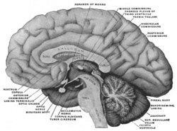 Mesal aspect of a brain sectioned in the median sagittal plane. | |
| Details | |
| Identifiers | |
| Latin | diencephalon |
| MeSH | A08.186.211.730.385 |
| Code | TH H3.11.03.5.00001 |
| NeuroNames | hier-271 |
| NeuroLex ID | Diencephalon |
| TA |
A14.1.03.007 A14.1.08.001 |
| FMA | 62001 |
The diencephalon is part of the prosencephalon (forebrain), which develops from the foremost primary cerebral vesicle. The prosencephalon differentiates into a caudal diencephalon and rostral telencephalon. The cerebral hemispheres develop from the sides of the telencephalon, each containing a lateral ventricle. The diencephalon consists of structures that are lateral to the third ventricle, and includes the thalamus, the hypothalamus, the epithalamus and the subthalamus.
Structure

The diencephalon consists of the following structures:
- Thalamus
- Hypothalamus including the Neurohypophysis
- Epithalamus which consists of
- Anterior and Posterior Paraventricular nuclei
- Medial and lateral Habenular nuclei
- Stria medullaris thalami
- Posterior commissure
- Pineal body
Attachments
The optic nerve (CNII) attaches to the diencephalon. The optic nerve is a sensory (afferent) nerve responsible for vision; it runs from the eye through the optic canal in the skull and attaches to the diencephalon. The retina itself is derived from the optic cup, a part of the embryonic diencephalon.
Function
The diencephalon is the region of the embryonic vertebrate neural tube that gives rise to posterior forebrain structures including the thalamus, hypothalamus, posterior portion of the pituitary gland, and pineal gland. The hypothalamus performs numerous vital functions, most of which relating directly or indirectly to the regulation of visceral activities by way of other brain regions and the autonomic nervous system.
Additional images
-
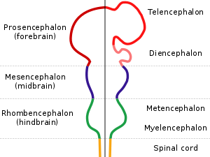
Diagram depicting the main subdivisions of the embryonic vertebrate brain. These regions will later differentiate into forebrain, midbrain and hindbrain structures.
-

Reconstruction of peripheral nerves of a human embryo of 10.2 mm. (Label for Diencephalon is at left.)
See also
References
This article incorporates text in the public domain from the 20th edition of Gray's Anatomy (1918)
External links
- Stained brain slice images which include the "diencephalon" at the BrainMaps project
- NIF Search - Diencephalon via the Neuroscience Information Framework