Dental extraction
| Dental extraction | |
|---|---|
| Intervention | |
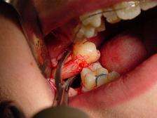 Surgical extraction of an impacted molar | |
| ICD-9-CM | 23.0-23.1 |
| MeSH | D014081 |


A dental extraction (also referred to as tooth extraction, exodontia, exodontics, or informally, tooth pulling) is the removal of teeth from the dental alveolus (socket) in the alveolar bone. Extractions are performed for a wide variety of reasons, but most commonly to remove teeth which have become unrestorable through tooth decay, periodontal disease or dental trauma, especially when they are associated with toothache. Sometimes wisdom teeth are impacted (stuck and unable to grow normally into the mouth) and may cause recurrent infections of the gum (pericoronitis). In orthodontics if the teeth are crowded, sound teeth may be extracted (often bicuspids) to create space so the rest of the teeth can be straightened.
Tooth extraction is usually relatively straightforward, and the vast majority can be usually performed quickly while the individual is awake by using local anesthetic injections to eliminate painful sensations. Local anesthetic blocks pain, but mechanical forces are still vaguely felt. Some teeth are more difficult to remove for several reasons, especially related to the tooth's position, the shape of the tooth roots and the integrity of the tooth. Dental phobia is an issue for some individuals, and tooth extraction tends to be feared more than other dental treatments like fillings. If a tooth is buried in the bone, a surgical or trans alveolar approach may be required, which involves cutting the gum away and removal of the bone which is holding the tooth in with a surgical drill. After the tooth is removed, stitches are used to replace the gum into the normal position.
Immediately after the tooth is removed, a bite pack is used to apply pressure to the tooth socket and stop the bleeding. After a tooth extraction, dentists usually give advice which revolves around not disturbing the blood clot in the socket by not touching the area with a finger or the tongue, by avoiding vigorous rinsing of the mouth and avoiding strenuous activity. Sucking, such as through a straw, is to be avoided. If the blood clot is dislodged, bleeding can restart, or alveolar osteitis ("dry socket") can develop, which can be very painful and lead to delayed healing of the socket. Smoking is avoided for at least 24 hours as it impairs wound healing and makes dry socket significantly more likely. Most advise hot salt water mouth baths which start 24 hours after the extraction.
The branch of dentistry that deals primarily with extractions is oral surgery ("exodontistry"), although general dentists and periodontists often carry out tooth extraction routinely since it is a core skill taught in dental schools. Periodontists are performing more and more extractions, since they often follow up and place a dental implant.
Reasons
The most common reason for extraction is tooth damage due to breakage or decay. There are additional reasons for tooth extraction:
- Severe tooth decay or infection (acute or chronic alveolar abscess[1]). Despite the reduction in worldwide prevalence of dental caries, it is still the most common reason for extraction of (non-third molar) teeth with up to two thirds of extractions.[2]
- Supernumerary teeth which are blocking other teeth from coming in.
- Severe gum disease which may affect the supporting tissues and bone structures of teeth.
- In preparation for orthodontic treatment (braces)
- Teeth in the fracture line
- Teeth which cannot be restored endodontically
- Fractured teeth
- Supernumerary, supplementary or malformed teeth
- Prosthetics; teeth detrimental to the fit or appearance of dentures[3]
- Treatment of symptomatic impacted wisdom teeth, whose impaction is causing pathosis that will lead to yet more (infection, inflammation, bone resorption)
- Preventive/prophylactic removal of asymptomatic impacted wisdom teeth. Although many dentists remove asymptomatic impacted third molars,[4][5] both American and British Health Authorities recommend against this routine procedure, unless there is evidence for disease in the impacted tooth or the near environment.[6] The American Public Health Association, for example, adopted a policy, Opposition to Prophylactic Removal of Third Molars (Wisdom Teeth), because of the large number of injuries resulting from unnecessary extractions.[7]
- Cosmetic - to remove teeth of poor appearance, unsuitable for restoration
- Head and neck radiation therapy, to treat and/or manage tumors, may require extraction of teeth, either before or after radiation treatments
- Deliberate, medically unnecessary, extraction as a form of physical torture.[8]
- It was once a common practice to remove the front teeth of institutionalized psychiatric patients who had a history of biting.[9]
- Reduced cost compared to other treatments[10]:98
Types
Extractions are often categorized as "simple" or "surgical".
Simple extractions are performed on teeth that are visible in the mouth, usually under local anaesthetic, and require only the use of instruments to elevate and/or grasp the visible portion of the tooth. Typically the tooth is lifted using an elevator, and using dental forceps, rocked back and forth until the periodontal ligament has been sufficiently broken and the supporting alveolar bone has been adequately widened to make the tooth loose enough to remove. Typically, when teeth are removed with forceps, slow, steady pressure is applied with controlled force.
Surgical extractions involve the removal of teeth that cannot be easily accessed, either because they have broken under the gum line or because they have not erupted fully. Surgical extractions almost always require an incision. In a surgical extraction the doctor may elevate the soft tissues covering the tooth and bone and may also remove some of the overlying and/or surrounding jawbone tissue with a drill or osteotome. Frequently, the tooth may be split into multiple pieces to facilitate its removal. Surgical extractions are usually performed under a general anaesthetic.
Anticoagulant use
Studies have shown that there is a correlation between consumption of anticoagulant drugs after dental extractions and the amount of bleeding. In one such review, oral anticoagulants were prescribed to multiple subjects, all of whom were undergoing dental surgery. 89 out of 990 subjects (9%) had delayed postoperative bleeding, and 3.5% of these cases were not controlled by local measures (‘serious cases’).[11] Other studies have reported greater numbers of patients with minor post-operative bleeding.[12] However, it is difficult to standardise bleeding as the definitions used to categorise the extent of the bleed tend to differ from study to study. However, the majority of studies concur that there is little risk of a major bleed if a patient is regularly consuming oral anticoagulants at the time of a simple dental extraction.
For simple extractions, therapeutic anticoagulation can be continued, as the bleeding risk is not high[13][14][15] and the risk of a thromboembolism caused by a temporary withdrawal from the anticoagulant is much higher than that of a serious bleed following the extraction[16] However, for complex extractions (3 or more teeth or multiple adjacent teeth), the risk of bleeding is higher,[17] and the dentist should consult the patient’s doctor. Patients undergoing a course of treatment using anticoagulants should notify their dentist when organising the procedure. An individual treatment plan should be drawn up for the patient, and the patient’s doctor should be contacted to confirm the anticoagulant being used, and the dose type.[17] The patient’s INR should also be taken into account. When the patient has an INR of 4.0 or over, they should be referred to a specialist [16] The risk of haemorrhage is increased in the elderly (especially after post-surgical dental extractions) as they are more susceptible to dental caries and periodontal diseases.[18] This should also be taken into account by the dentist.
To increase the effectiveness of oral anticoagulant drugs, bleeding risks can be further minimized by the usage of collagen sponges and sutures and rinsing 5% tranexamic acid mouthwash four times a day.[18]
Overall, patients utilizing long-term anticoagulant therapies such as warfarin or salicylic acid do not need to discontinue its use prior to having a tooth extracted. The extraction should be performed utilizing the least traumatic extraction procedures[19] and patients should make sure to tell their dentist or oral surgeon about any medications they may take before the procedure.
Antibiotic use
Antibiotics can be prescribed by dental professionals to reduce risks of certain post extraction complications. There is evidence that use of antibiotics before and/or after impacted wisdom tooth extraction reduces the risk of infections by 70% and lowers incidence of dry socket by one third. For every 12 people who are treated with an antibiotic following impacted wisdom tooth removal, one infection is prevented. Use of antibiotics does not seem to have a direct effect on manifestation of fever, swelling or trismus seven days post-extraction. In the 2013 Cochrane review, 18 randomized control double-blinded experiments were reviewed and after considering the biased risk associated with these studies, it was concluded that there is moderate overall evidence supporting the routine use of antibiotics in practice in order to reduce risk of infection following a third molar extraction. There are still reasonable concerns remaining regarding the possible adverse effects of indiscriminate antibiotic use in patients. There are also concerns about development of antibiotic resistance which advises against the use of prophylactic antibiotics in practice.[20]
Post-extraction healing

Immediately following the removal of a tooth, bleeding or just oozing very commonly occurs. Pressure is applied by biting on a gauze swab, and a thrombus (blood clot) forms in the socket (hemostatic response). Common haemostatic measures include local pressure application with gauze and the used of oxidized cellulose (gelfoam) and fibrin sealant. Dental practitioners usually have absorbent gauze, haemostatic packing material (oxidised cellulose, collagen sponge) and suture kit available.[21] Sometimes 30 minutes of continuous pressure is required to fully arrest bleeding. Talking, which moves the mandible and hence removes the pressure applied on the socket, instead of keeping constant pressure, is a very common reason that bleeding might not stop. This is likened to someone with a bleeding wound on their arm, when being instructed to apply pressure, instead holds the wound intermittently every few moments. Coagulopathies (clotting disorders, e.g. hemophilia) are sometimes discovered for the first time if a person has had no other surgical procedure in their life, but this is rare. Sometimes the blood clot can be dislodged, triggering more bleeding and formation of a new blood clot, or leading to a dry socket (see complications). Some oral surgeons routinely scrape the walls of a socket to encourage bleeding in the belief that this will reduce the chance of dry socket, but there is no evidence that this practice works.
The chance of further bleeding reduces as healing progresses, and is unlikely after 24 hours. If the bleeding occurs beyond 8 –12 hours, this situation is
then referred as post-extraction bleeding. The blood clot is covered by epithelial cells which proliferate from the gingival mucosa of socket margins, taking about 10 days to fully cover the defect.[22] In the clot, neutrophils and macrophages are involved as an inflammatory response takes place. The proliferative and synthesizing phase next occurs, characterized by proliferation of osteogenic cells from the adjacent bone marrow in the alveolar bone. Bone formation starts after about 10 days from when the tooth was extracted. After 10–12 weeks, the outline of the socket is no longer apparent on an X-ray image. Bony remodeling as the alveolus adapts to the edentulous state occurs in the longer term as the alveolar process slowly resorbs. In maxillary posterior teeth, the degree of pneumatization of the maxillary sinus may also increase as the antral floor remodels.
Post Extraction Bleeding[23]
Post extraction bleeding is bleeding that occurs 8–12 hours after tooth extraction. There are various factors that contribute to post-extraction bleeding.
Local factors
- Laceration of blood vessels
- Osseous bleeding from nutrients canal/ central vessels
- Inflammation
- Infection
- Traumatic extraction
- Failure of patients to follow post-extraction instruction
Systemic factors
- Platelet problem
- Coagulation disorder/ excessive fibrinolysis
- Inherited/ medication-induced problems
Type of Bleeding
1. Primary prolonged bleeding
This type of bleeding occurs during/ immediately after extraction, due to infection or trauma to blood vessels. It is usually controlled by conventional techniques such as applying pressure packs or haemostatic agents onto the wound.
2. Reactionary bleeding
This type of bleeding starts 2 to 3 hours after tooth extraction, due to systemic conditions. Systemic intervention might be required.
3. Secondary bleeding
This type of bleeding usually begins 7 to 10 days post extraction and is said to be rare. It happens mainly due to secondary infection.
Interventions
When dental practitioner is deciding on how to control post-extraction bleeding, many factors have to be taken into account:
- Surgical area
- Location of bleeding
- Size of wound
- Extend of bleeding
- Accessibility of bleeding site
- Time of bleeding
Post-extraction bleeding interventions can be categorized into two main groups:
Local interventions
(i) Surgical interventions
- Involve suturing the bleeding site
(ii) Non-surgical haemostatic measures
- Involve the use of drugs, sealants, adhesives, absorbable agents, biologics, and combination of products
(iii) Combination of both
2. Systemic interventions
This is important for patients who have systemic cause for bleeding. Usually, local haemostatics do not work well on limiting their bleeding because they only result in temporary cessation of bleeding.
Complications

- Infection: The dentists may opt to prescribe antibiotics pre- and/or post-operatively if they determine the patient to be at risk.
- Prolonged bleeding: The dentist has a variety of means at their disposal to address bleeding; however, small amounts of blood mixed in the saliva after extractions are normal, even up to 72 hours after extraction. Usually, however, bleeding will almost completely stop within eight hours of the surgery, with only minuscule amounts of blood mixed with saliva coming from the wound. A gauze compress will significantly reduce bleeding over a period of a few hours.
- Swelling: Often dictated by the amount of surgery performed to extract a tooth (e.g. surgical insult to the tissues both hard and soft surrounding a tooth). Generally, when a surgical flap must be elevated (i.e. and the periosteum covering the bone is thus injured), minor to moderate swelling will occur. A poorly cut soft tissue flap, for instance, where the periosteum is torn off rather than cleanly elevated off the underlying bone, will often increase such swelling. Similarly, when bone must be removed using a drill, more swelling is likely to occur.
- Bruising: Bruising may occur as a complication after tooth extraction. Bruising is more common in older people or people on aspirin or steroid therapy. It may take weeks for bruising to disappear completely.
- Sinus exposure and oral-antral communication: This can occur when extracting upper molars (and in some patients, upper premolars). The maxillary sinus sits right above the roots of maxillary molars and premolars. There is a bony floor of the sinus dividing the tooth socket from the sinus itself. This bone can range from thick to thin from tooth to tooth from patient to patient. In some cases it is absent and the root is in fact in the sinus. At other times, this bone may be removed with the tooth, or may be perforated during surgical extractions. The doctor typically mentions this risk to patients, based on evaluation of radiographs showing the relationship of the tooth to the sinus. The sinus cavity is lined with a membrane called the Sniderian membrane, which may or may not be perforated. If this membrane is exposed after an extraction, but remains intact, a "sinus exposed" has occurred. If the membrane is perforated, however, it is a "sinus communication". These two conditions are treated differently. In the event of a sinus communication, the dentist may decide to let it heal on its own or may need to surgically obtain primary closure—depending on the size of the exposure and the likelihood of the patient to heal. In both cases, a resorbable material called "gelfoam" is typically placed in the extraction site to promote clotting and serve as a framework for granulation tissue to accumulate. Patients are typically provided with prescriptions for antibiotics that cover sinus bacterial flora, decongestants, and careful instructions to follow during the healing period.
- Nerve injury: This is primarily an issue with extraction of third molars, but can occur with the extraction of any tooth should the nerve be close to the surgical site. Two nerves are typically of concern, and are found in duplicate (one left and one right): 1. the inferior alveolar nerve, which enters the mandible at the mandibular foramen and exits the mandible at the sides of the chin from the mental foramen. This nerve supplies sensation to the lower teeth on the right or left half of the dental arch, as well as sense of touch to the right or left half of the chin and lower lip. 2. The lingual nerve (one right and one left), which branches off the mandibular branches of the trigeminal nerve and courses just inside the jaw bone, entering the tongue and supplying sense of touch and taste to the right and left half of the anterior 2/3 of the tongue as well as the lingual gingiva (i.e. the gums on the inside surface of the dental arch). Such injuries can occur while lifting teeth (typically the inferior alveolar), but are most commonly caused by inadvertent damage with a surgical drill. Such injuries are rare and are usually temporary, but depending on the type of injury (i.e. Seddon classification: neuropraxia, axonotmesis, & neurotmesis), can be prolonged or even permanent.
- Displacement of tooth or part of the tooth into the maxillary sinus (upper teeth only). In such cases, the tooth or tooth fragment must almost always be retrieved. In some cases, the sinus cavity can be irrigated with saline (antral lavage) and the tooth fragment may be brought back to the site of the opening through which it entered the sinus, and may be retrievable. At other times, a window must be made into the sinus in the Canine fossa—a procedure referred to as a "Caldwell-Luc".
- Dry socket (Alveolar osteitis) is a painful phenomenon that most commonly occurs a few days after the removal of mandibular (lower) wisdom teeth. It typically occurs when the blood clot within the healing tooth extraction site is disrupted. More likely, alveolar osteitis is a phenomenon of painful inflammation within the empty tooth socket because of the relatively poor blood supply to this area of the mandible (which explains why dry socket is usually not experienced in other parts of the jaws). Inflamed alveolar bone, unprotected and exposed to the oral environment after tooth extraction, can become packed with food and debris. A dry socket typically causes a sharp and sudden increase in pain commencing 2–5 days following the extraction of a mandibular molar, most commonly the third molar.[24] This is often extremely unpleasant for the patient; the only symptom of dry socket is pain, which often radiates up and down the head and neck. A dry socket is not an infection, and is not directly associated with swelling because it occurs entirely within bone – it is a phenomenon of inflammation within the bony lining of an empty tooth socket. Because dry socket is not an infection, the use of antibiotics has no effect on its rate of occurrence. There is some evidence that rinsing with chlorhexidine before or after extraction or placing chlorhexidine gel in the sockets of extracted teeth provides a benefit in preventing dry socket, but potential adverse effects of chlorhexidine have to be considered.[25] The risk factor for alveolar osteitis can dramatically increase with smoking after an extraction.
- Bone fragments: Particularly when extraction of molars is involved, it is not uncommon for the bones which formerly supported the tooth to shift and in some cases to erupt through the gums, presenting protruding sharp edges which can irritate the tongue and cause discomfort. This is distinguished from a similar phenomenon where broken fragments of bone or tooth left over from the extraction can also protrude through the gums. In the latter case, the fragments will usually work their way out on their own. In the former case, the protrusions can either be snipped off by the dentist, or eventually the exposed bone will erode away on its own.
- Trismus: Trismus, also known as lockjaw, affects functions of the oral cavity by restricting opening of the mouth. A double blind, clinical study was done to test the effect of two different medications on post-extraction trismus. The patients who received a corticosteroid by IV had a statistically significant lower level of trismus when compared to patients receiving an NSAID by IV or no medication.[26]
- Loss of a tooth: If an extracted tooth slips out of the forceps, it may be swallowed or inhaled. The patient may be aware of swallowing it, or they may cough, which suggests inhalation of the tooth. The patient must be referred to for a chest X-ray in hospital if a tooth cannot be found. If it has been swallowed, no action is necessary as it usually passes through the alimentary canal without doing any harm. But if it has been inhaled, an urgent operation is necessary to recover it from the airway or lung before it causes serious complications such as pneumonia or a lung abscess.[3]
- Luxation of the adjacent tooth: The force application during extraction procedure must strictly be limited to the tooth that requires the extraction. Most case of surgical extraction procedures require that the forces are diverted from the tooth itself to areas such as bone surrounding the tooth to ensure adequate bone removal before proceeding any further in the extraction procedure. Either ways the forces applied by various instruments during both simple and complicated surgical procedure may loosen the teeth present both in-front or behind of the tooth depending upon the impact, direction and location of the force being applied, and that happening only if the forces divert from the actual tooth that needs extraction. Such deleterious forces may weaken the anchorage of adjacent teeth from within their boney socket and hence result in weakening of the adjacent teeth.
- Extraction of the wrong tooth: Misdiagnosis, altered tooth morphology, faulty clinical examination, poor patient history, undetected / unmentioned previous extractions that may predispose the operator to consider another tooth to be a replicate of the one previously extracted are a few causes of extraction of a wrong tooth.
- Osteonecrosis: Osteonecrosis of the jaw is the slow destruction of bone in an extraction site. A case control study of 191 cases and 573 controls were used to understand the relationship between osteonecrosis of the jaw and prior usage of bisphosphonate drugs, which are commonly prescribed to treat osteoporosis. All of the participants were over 40 years of age, mostly female, and had been taking bisphosphonates for six months or longer. The presence of osteonecrosis of the jaw was reported by dentists' previous diagnosis of the participating case and control patient's medical records. Reports showed that women using bisphosphonates for more than two years are ten times more likely to experience osteonecrosis of the jaw, while women who have taken bisphosphonates for less than two years are four times more likely to suffer from osteonecrosis of the jaw when compared to women who were not taking bisphosphonates. Therefore, it is extremely important to report all medications used to the dentist before an extraction, so that osteonecrosis can be avoided.[27]
Assessing nerve injury risk
There are specific factors that need to be accounted for when considering nerve injury after removal of mandibular third molars (bottom wisdom teeth). Position of the molars is an important risk factor with regards to inferior alveolar nerve injuries. Horizontally-impacted molars pose a higher risk of nerve injury, as the depth of the impacted molar is increased. Furthermore, the most important factor for inferior alveolar nerve-injury prediction is the proximity of the root tips to the mandibular canal.[28]
Pain management
Many drug therapies are available for pain management after third molar extractions including NSAIDS (non-steroidal anti-inflammatory), APAP (acetaminophen) and opioid formulations. Although each has its own pain relieving efficacy, they also pose adverse effects. According to Dr. Paul A Moore and Dr. Elliot V. Hersh, Ibuprofen-APAP combinations have the greatest efficacy in pain relief and reducing inflammation along with the fewest adverse effects. Taking either of these agents alone or in combination may be contraindicated in those who have certain medical conditions. For example, taking ibuprofen or any NSAID in conjunction with warfarin (a blood thinner) may not be appropriate. Also, prolonged use of ibuprofen or APAP has GI and cardiovascular risks.[29]
Socket preservation
Socket preservation or alveolar ridge preservation (ARP)[30] is a procedure to reduce bone loss after tooth extraction to preserve the dental alveolus (tooth socket) in the alveolar bone. At the time of extraction a platelet rich fibrin (PRF)[31] membrane containing bone growth enhancing elements is placed in the wound or a graft material or scaffold is placed in the socket of the extracted tooth.[32][33] The socket is then directly closed with stitches or covered with a non-resorbable or resorbable membrane and sutured.[34]
Replacement options for missing teeth
Following dental extraction, a gap is left. The options to fill this gap are commonly recorded as "BIND", and the exact choice is agreed between dentist and patient based upon several factors.
| Treatment option | Advantages | Disadvantages |
|---|---|---|
| Bridge | Fixed to adjacent teeth | Drilling usually required on one or both sides of the gap if conventional bridge (average lifespan about 10 years). Conservative bridge (average lifespan about 5 years) preparation may cause minimal damage to adjacent teeth. Expensive and complex treatment, not suited to all situations, e.g. large gaps in the back of the mouth Alveolar bone will still resorb, and eventually a gap may show under bridge. |
| Implant | Fixed to jawbone. Maintains alveolar bone, which would otherwise undergo resorption. Usually a long term lifespan. | Expensive and complex, requiring specialist. May involve other procedures such as bone grafting. Relatively contra-indicated in tobacco smokers. |
| Nothing (i.e. not replacing the missing tooth) | Often the choice due to cost of other treatment or lack of motivation for other treatments. Part of a shortened dental arch plan, which revolves around the fact that not all teeth are required to eat comfortably, and only the incisors and premolars need be preserved for normal function. | The alveolar bone will slowly resorb over time once the tooth is lost. Potential esthetic concern. Potential for drifting and rotation of adjacent teeth into the gap over time. |
| Denture | Often a simple, quick and relatively cheap treatment compared to bridge and implant. Not usually any drilling of other teeth required. It is far easier to replace several teeth with a denture than place multiple bridges or implants. | Denture is not fixed in mouth. Over time worsens periodontal disease unless there is good level of oral hygiene, and may damage soft tissues. Potential for slightly accelerated resorption of alveolar bone compared to no denture. Potential for poor tolerance in persons with over-sensitive gag reflex, xerostomia, etc. |
History
Historically, dental extractions have been used to treat a variety of illnesses. Before the discovery of antibiotics, chronic tooth infections were often linked to a variety of health problems, and therefore removal of a diseased tooth was a common treatment for various medical conditions. Instruments used for dental extractions date back several centuries. In the 14th century, Guy de Chauliac invented the dental pelican,[35] which was used through the late 18th century. The pelican was replaced by the dental key which, in turn, was replaced by modern forceps in the 20th century. As dental extractions can vary tremendously in difficulty, depending on the patient and the tooth, a wide variety of instruments exist to address specific situations. Rarely, tooth extraction was used as a method of torture, e.g. to obtain forced confessions.[36]
See also
References
- ↑ Kazemi, Dr. H. Ryan (2014), EXTRACTION OF DAMAGED TEETH, retrieved February 18, 2014
- ↑ Zadik Y, Sandler V, Bechor R, Salehrabi R (August 2008). "Analysis of factors related to extraction of endodontically treated teeth". Oral Surg Oral Med Oral Pathol Oral Radiol Endod. 106 (5): e31–5. doi:10.1016/j.tripleo.2008.06.017. PMID 18718782.
- 1 2 Hollins, Carole (2008). Levison's Textbook for Dental Nurses. ISBN 978-1-4051-7557-9
- ↑ Zadik Y, Levin L (April 2006). "[Decision making of Hebrew University and Tel Aviv University Dental Schools graduates in every day dentistry--is there a difference?]". Refuat Hapeh Vehashinayim (in Hebrew). 23 (2): 19–23, 65. PMID 16886872.
- ↑ Zadik Y, Levin L (April 2007). "Decision making of Israeli, East European, and South American dental school graduates in third molar surgery: is there a difference?". J. Oral Maxillofac. Surg. 65 (4): 658–62. doi:10.1016/j.joms.2006.09.002. PMID 17368360.
- ↑ Morant H (April 200). "UK government wants GMC to be given stronger powers". British Medical Journal. 320 (7239): 890A. doi:10.1136/bmj.320.7239.890. PMC 1117823
 . PMID 10741982. Retrieved 2008-08-08. Check date values in:
 |date=(help) - ↑ "Opposition to Prophylactic Removal of Third Molars (Wisdom Teeth)". Policy Statement Database. American Public Health Association. 2016-05-12. Retrieved 2016-05-12.
- ↑ Speers, RD; Brands, WG; Nuzzolese, E; Smith, D; Swiss, PB; van Woensel, M; Welie, JV (Dec 2008). "Preventing dentists' involvement in torture: the developmental history of a new international declaration.". Journal of the American Dental Association (1939). 139 (12): 1667–73. doi:10.14219/jada.archive.2008.0109. PMID 19047673.
- ↑ C. Thomas Gualtieri. Brain injury and mental retardation: psychopharmacology and neuropsychiatry.
- ↑ Hupp JR, Ellis E, Tucker MR (2008). Contemporary oral and maxillofacial surgery (5th ed.). St. Louis, Mo.: Mosby Elsevier. ISBN 9780323049030.
- ↑ Randall, C (2005). "Surgical Management of the Primary Care Dental Patient on Warfarin". Dental Update. 32: 414–424.
- ↑ Al-Mubarak, S (2006). "Thromboembolic Risk and Bleeding in Patients Maintaining or Stopping Oral Anticoagulant Therapy During Dental Extraction". J Thromb Haemost. 4: 689–691.
- ↑ Abdullah, Walid Ahmed; Khalil, Hesham (2014-08-19). "Dental extraction in patients on warfarin treatment". Clinical, Cosmetic and Investigational Dentistry. 6: 65–69. doi:10.2147/CCIDE.S68641. ISSN 1179-1357. PMC 4144934. PMID 25170281.
- ↑ "Guidelines | British Society for Haematology" (PDF). www.bcshguidelines.com. Retrieved 2016-11-22.
- ↑ "UC Davis Health System Anticoagulation Services Recommendations for anticoagulation management before and after dental procedures" (PDF). UCDHS Pharmacy and Therapeutics Committee. 2015.
- 1 2 "Managing patients who are taking warfarin and undergoing dental treatment". NHS Guidelines. NHS, British Dental Association and advice from the Haemostasis and Thrombosis Task Force of the British Committee for Standards in Haematology (BCSH). 2004.
- 1 2 "Management of Dental Patients Taking Anticoagulants or Antiplatelet Drugs - Dental Clinical Guidance" (PDF). Scottish Dental Clinical Effectiveness Programme. 2015.
- 1 2 Yang, Shuo; Shi, Quan; Liu, Jinglong; Li, Jinru; Xu, Juan (2016-01-01). "Should oral anticoagulant therapy be continued during dental extraction? A meta-analysis". BMC Oral Health. 16: 81. doi:10.1186/s12903-016-0278-9. ISSN 1472-6831. PMC 5002166. PMID 27566540.
- ↑ Perieira, CM (2011). "Tooth extraction in patients on oral anticoagulants: prospective study conducted in 108 brazilian patients". ISRN Dent. 2011: 203619. doi:10.5402/2011/203619. PMC 3169237. PMID 21991458.
- ↑ Lodi, G (Nov 2012). "Antibiotics to prevent complications following tooth extractions". Cochrane Database Syst Rev. 11: CD003811. doi:10.1002/14651858.CD003811.pub2. PMID 23152221.
- ↑ "Management of Dental Patients taking Anticoagulant or Anti Platelet Drug" (PDF).
- ↑ Antonio Nanci (editor) (2007). Oral histology : development, structure, and function. (7th ed.). St. Louis, Mo.: Mosby. ISBN 9780323045575.
- ↑ Sumanth, Kumbargere N; Prashanti, Eachempati; Aggarwal, Himanshi; Kumar, Pradeep; Lingappa, Ashok; Muthu, Murugan S; Kiran Kumar Krishanappa, Salian (2016-06-10). Cochrane Database of Systematic Reviews. John Wiley & Sons, Ltd. doi:10.1002/14651858.cd011930.pub2.
- ↑ Daly, B; et al. (Dec 12, 2012). "Local interventions for the management of alveolar osteitis (dry socket)". Cochrane Database Sys Rev. 12: CD006968. doi:10.1002/14651858.CD006968.pub2. PMID 23235637.
- ↑ Dodson, T (Mar 2013). "Prevention and treatment of dry socket". Evid Based Dent. 1 (14): 13–4. doi:10.1038/sj.ebd.6400913. PMID 23579300.
- ↑ Ilhan, O; et al. (2014). "A comparison of the effects of methylprednisolone and tenoxicam on pain, edema, and trismus after impacted lower third molar extraction". Medical Science Monitor: International Medical Journal of Experimental and Clinical Research. 20: 147–152. doi:10.12659/MSM.890239. PMC 3915002. PMID 24473372.
- ↑ Barasch, A (April 2011). "Risk factors for osteonecrosis of the jaws: A case-control study from the CONDOR dental PBRN". J Dent Res. 90 (4): 439–44.
- ↑ Juodzbalys, G (Jul 1, 2013). "Manibular Third Molar Impaction: Review of the Literature and a Proposal of a Classification". J Oral Maxillofac Res. 4 (2): e1. doi:10.5037/jomr.2013.4201. PMC 3886113. PMID 24422029.
- ↑ Moore, PA (Aug 2013). "COmbining ibuprofen and acetaminophen for acute pain management after third molar extractions: translating clinical research to d". J Am Dent Assoc. 144 (8): 898–908. doi:10.14219/jada.archive.2013.0207. PMID 23904576.
- ↑ Peck, Mogammad Thabit; Marnewick, Johan; Stephen, Lawrence (2011). "Alveolar Ridge Preservation Using Leukocyte and Platelet-Rich Fibrin: A Report of a Case". Case Reports in Dentistry. 2011: 1–5. doi:10.1155/2011/345048. ISSN 2090-6447.
- ↑ Khiste, Sujeet Vinayak; Naik Tari, Ritam (2013). "Platelet-Rich Fibrin as a Biofuel for Tissue Regeneration". ISRN Biomaterials. 2013: 1–6. doi:10.5402/2013/627367. ISSN 2314-4025.
- ↑ Tassos Irinakis, Rationale for Socket Preservation after Extraction of a Single-Rooted Tooth when Planning for Future Implant Placement, Journal of Canadian Dental Association 2006; 72(10):917–922
- ↑ Fickl, Stefan; Zuhr, Otto; Wachtel, Hannes; Stappert, Christian F. J.; Stein, Jamal M.; Hürzeler, Markus B. (2008). "Dimensional changes of the alveolar ridge contour after different socket preservation techniques". Journal of Clinical Periodontology. 35 (10): 906–913. doi:10.1111/j.1600-051X.2008.01305.x. ISSN 0303-6979.
- ↑ Extraction socket preservation: The time is now
- ↑ "Dental pelican for tooth pulling, Europe, 1701–1800". sciencemuseum.org.uk. Brought to Life. Retrieved 18 February 2014.
- ↑ Downs, edited by Richard Pierre Claude and Burns H. Weston, with the assistance of Gavin H. Boyles and Jessica L. (2006). Human rights in the world community : issues and action (3rd ed.). Philadelphia: University of Pennsylvania Press. p. 91. ISBN 9780812219487.