Deep cervical fascia
| Deep cervical fascia | |
|---|---|
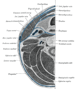 Section of the neck at about the level of the sixth cervical vertebra. Showing the arrangement of the fascia coli. | |
The deep cervical fascia (or fascia colli in older texts) lies under cover of the Platysma, and invests the neck; it also forms sheaths for the carotid vessels, and for the structures situated in front of the vertebral column. Its attachment to the hyoid bone prevents the formation of a dewlap.[1]
The investing portion of the fascia is attached behind to the ligamentum nuchæ and to the spinous process of the seventh cervical vertebra.
The alar fascia is a portion of the deep cervical fascia.
Divisions
The deep cervical fascia is often divided into a superficial, middle, and deep layer.
The superficial layer envelopes the trapezius, sternocleidomastoid, and muscles of facial expression. It also contains the submandibular and parotid salivary gland as well as the muscles of mastication (the masseter, pterygoid, and temporalis muscles).
The middle layer envelopes the strap muscles (sternohyoid, sternothyroid, thyrohyoid, and omohyoid muscles). It also surrounds the pharynx, larynx, trachea, esophagus, thyroid, parathyroids, buccinators, and constrictor muscles of the pharynx.
The deep layer is the pre vertebral fascial layer and surrounds the paraspinous muscles and cervical vertebrae.[2]
Superior attachments and relations
Above, the fascia is attached to the superior nuchal line of the occipital bone, to the mastoid process of the temporal bone, and to the whole length of the inferior border of the body of the mandible.
Opposite the angle of the mandible the fascia is very strong, and binds the anterior edge of the Sternocleidomastoideus firmly to that bone.
Between the mandible and the mastoid process it ensheathes the parotid gland—the layer which covers the gland extends upward under the name of the parotideomasseteric fascia and is fixed to the zygomatic arch.
From the part which passes under the parotid gland a strong band extends upward to the temporal styloid process, forming the stylomandibular ligament.
Two other bands may be defined: the sphenomandibular and the pterygospinous ligaments.
The pterygospinous ligament stretches from the upper part of the posterior border of the lateral pterygoid plate to the spinous process of the sphenoid.
It occasionally ossifies, and in such cases, between its upper border and the base of the skull, a foramen is formed which transmits the branches of the mandibular nerve to the muscles of mastication.
Inferior attachments and relations
Below, the fascia is attached to the acromion, the clavicle, and the manubrium sterni.
Some little distance above the last it splits into two layers, superficial and deep.
The former is attached to the anterior border of the manubrium, the latter to its posterior border and to the interclavicular ligament.
Between these two layers is a slit-like interval, the suprasternal space (space of Burns); it contains a small quantity of areolar tissue, the lower portions of the anterior jugular veins and their transverse connecting branch, the sternal heads of the Sternocleidomastoidei, and sometimes a lymph gland.
Processes
The fascia which lines the deep surface of the Sternocleidomastoideus gives off the following processes:
- A process envelops the tendon at the Omohyoideus, and binds it down to the sternum and first costal cartilage.
- A strong sheath, the carotid sheath, encloses the carotid artery, internal jugular vein, and vagus nerve.
- The prevertebral fascia extends medialward behind the carotid vessels, where it assists in forming their sheath, and passes in front of the prevertebral muscles.
- The pretracheal fascia extends medially in front of the carotid vessels, and assists in forming the carotid sheath.
References
This article incorporates text in the public domain from the 20th edition of Gray's Anatomy (1918)
External links
- Sagittal Section Showing Deep Cervical Fascial Layers
- Infrahyoid Cross-Section Showing Layers of Deep Cervical Fascia
- Suprahyoid Cross-Section Showing Layers of Deep Cervical Fascia