Color blindness
| Color blindness | |
|---|---|
| color deficiency, impaired color vision[1] | |
|
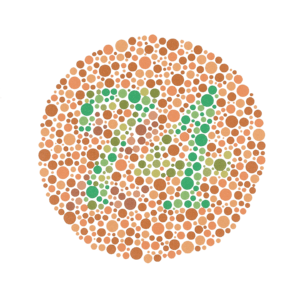
Example of an Ishihara color test plate. Depending on the computer displays, people with normal vision should see the number "74". Many people who are color blind see it as "21", and those with total color blindness may not see any numbers. | |
| Classification and external resources | |
| Specialty | Ophthalmology |
| ICD-10 | H53.5 |
| ICD-9-CM | 368.5 |
| DiseasesDB | 2999 |
| MedlinePlus | 001002 |
| MeSH | D003117 |
Color blindness, also known as color vision deficiency, is the decreased ability to see color or differences in color.[2] Color blindness can make some educational activities difficult. Buying fruit, picking clothing, and reading traffic lights can also be more challenging. Problems, however, are generally minor and most people adapt. People with total color blindness may also have decreased visual acuity and be uncomfortable in bright environments.[2]
The most common cause of color blindness is a fault in the development of one or more of the three sets of color sensing cones in the eye. Males are more likely to be color blind than females as the genes responsible for the most common forms of color blindness are on the X chromosome. As females have two X chromosomes, a defect in one is typically compensated for by the other, while males only have one X chromosome. Color blindness can also result from physical or chemical damage to the eye, optic nerve, or parts of the brain. Diagnosis is typically with the Ishihara color test; however a number of other testing methods also exist.[2]
There is no cure for color blindness.[2] Diagnosis may allow a person's teacher to change their method of teaching to accommodate the decreased ability to recognize color.[1] Special lenses may help people with red–green color blindness when under bright conditions. There are also mobile apps that can help people identify colors.[2]
Red–green color blindness is the most common form, followed by blue–yellow color blindness and total color blindness.[2] Red–green color blindness affects up to 8% of males and 0.5% of females of Northern European descent. The ability to see color also decreases in old age.[2] Being color blind may make people ineligible for certain jobs in certain countries. This may include pilot, train driver, and armed forces. The effect of color blindness on artistic ability, however, is controversial. The ability to draw appears to be unchanged and a number of famous artists are believed to have been color blind.[1]
Signs and symptoms
In almost all cases, color blind people retain blue–yellow discrimination, and most color-blind individuals are anomalous trichromats rather than complete dichromats. In practice, this means that they often retain a limited discrimination along the red–green axis of color space, although their ability to separate colors in this dimension is severely reduced. Color blindness very rarely means complete monochromatism.
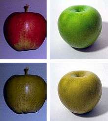
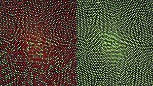
Dichromats often confuse red and green items. For example, they may find it difficult to distinguish a Braeburn apple from a Granny Smith and in some cases, the red and green of traffic lights without other clues—for example, shape or position. The vision of dichromats may also be compared to images produced by a color printer that has run out of the ink in one of its three color cartridges (for protanopes and deuteranopes, the red cartridge, and for tritanopes, the yellow cartridge). Dichromats tend to learn to use texture and shape clues and so are often able to penetrate camouflage that has been designed to deceive individuals with color-normal vision.[3]
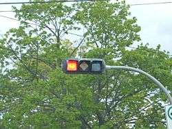
Colors of traffic lights are confusing to some dichromats, as there is insufficient apparent difference between the red/amber traffic lights, and that of sodium street lamps; also, the green can be confused with a grubby white lamp. This is a risk factor on high-speed undulating roads where angular cues cannot be used. British Rail color lamp signals use more easily identifiable colors: The red is blood red, the amber is yellow and the green is a bluish color. Most British road traffic lights are mounted vertically on a black rectangle with a white border (forming a "sighting board") and so dichromats can look for the position of the light within the rectangle—top, middle or bottom. In the eastern provinces of Canada horizontally mounted traffic lights are generally differentiated by shape to facilitate identification for those with color blindness. In the United States, this is not done by shape but by position, as the red light is always on the left if the light is horizontal, or on top if the light is vertical. However, a single flashing light (red indicating cars must stop, yellow for caution/yield) is indistinguishable, but these are rare.
-

Normal sight
-

Deuteranopia sight
-

Tritanopia sight
-

Monochromacy sight
Causes
Color vision deficiencies can be classified as acquired or inherited.
- Acquired: Diseases, drugs (e.g., Plaquenil), and chemicals may cause color blindness.[4][5]
- Inherited: There are three types of inherited or congenital color vision deficiencies: monochromacy, dichromacy, and anomalous trichromacy.
- Monochromacy, also known as "total color blindness", is the lack of ability to distinguish colors (and thus the person views everything as if it were on a black and white television); caused by cone defect or absence. Monochromacy occurs when two or all three of the cone pigments are missing and color and lightness vision is reduced to one dimension.
- Rod monochromacy (achromatopsia) is an exceedingly rare, nonprogressive inability to distinguish any colors as a result of absent or nonfunctioning retinal cones. It is associated with light sensitivity (photophobia), involuntary eye oscillations (nystagmus), and poor vision.
- Cone monochromacy is a rare total color blindness that is accompanied by relatively normal vision, electroretinogram, and electrooculogram. Cone monochromacy can also be a result of having more than one type of dichromatic color blindness. People who have, for instance, both protanopia and tritanopia are considered to have cone monochromacy. Since cone monochromacy is the lack of/damage of more than one cone in retinal environment, having two types of dichromacy would be an equivalent.
- Dichromacy is a moderately severe color vision defect in which one of the three basic color mechanisms is absent or not functioning. It is hereditary and, in the case of protanopia or deuteranopia, sex-linked, affecting predominantly males. Dichromacy occurs when one of the cone pigments is missing and color is reduced to two dimensions. Dichromacy conditions are labeled based on whether the "first" (Greek: prot-, referring to the red photoreceptors), "second" (deuter-, the green), or "third" (trit-, the blue) photoreceptors are affected.
- Protanopia is a severe type of color vision deficiency caused by the complete absence of red retinal photoreceptors. Protans have difficulties distinguishing between blue and green colors and also between red and green colors. It is a form of dichromatism in which the subject can only perceive light wavelengths from 400 to 650 nm, instead of the usual 700 nm. Pure reds cannot be seen, instead appearing black; purple colors cannot be distinguished from blues; more orange-tinted reds may appear as very dim yellows, and all orange–yellow–green shades of too long a wavelength to stimulate the blue receptors appear as a similar yellow hue. It is hereditary, sex-linked, and present in 1% of males.
- Deuteranopia is a type of color vision deficiency where the green photoreceptors are absent. It affects hue discrimination in the same way as protanopia, but without the dimming effect. Like protanopia, it is hereditary, sex-linked, and found in about 1% of the male population.[6]
- Tritanopia is a very rare color vision disturbance in which there are only two cone pigments present and a total absence of blue retinal receptors. Blues appear greenish, yellows and oranges appear pinkish, and purple colors appear deep red. It is related to chromosome 7. Unlike protanopia and deuteranopia, tritanopia and tritanomaly are not sex-linked traits and can be acquired rather than inherited and can be reversed in some cases.
- Anomalous trichromacy is a common type of inherited color vision deficiency, occurring when one of the three cone pigments is altered in its spectral sensitivity.
- Protanomaly is a mild color vision defect in which an altered spectral sensitivity of red retinal receptors (closer to green receptor response) results in poor red–green hue discrimination. It is hereditary, sex-linked, and present in 1% of males. The difference with protanopia is that in this case the L-cone is present but malfunctioning, whereas in the earlier the L-cone is completely missing.[7]
- Deuteranomaly, caused by a similar shift in the green retinal receptors, is by far the most common type of color vision deficiency, mildly affecting red–green hue discrimination in 5% of European males. It is hereditary and sex-linked. The difference with deuteranopia is that in this case the green sensitive cones are not missing but malfunctioning.[8]
- Tritanomaly is a rare, hereditary color vision deficiency affecting blue–green and yellow–red/pink hue discrimination. It is related to chromosome "7".[9] The difference is that the S-cone is malfunctioning but not missing.[10]
- Monochromacy, also known as "total color blindness", is the lack of ability to distinguish colors (and thus the person views everything as if it were on a black and white television); caused by cone defect or absence. Monochromacy occurs when two or all three of the cone pigments are missing and color and lightness vision is reduced to one dimension.
Genetics
Color blindness is typically inherited. It is most commonly inherited from mutations on the X chromosome but the mapping of the human genome has shown there are many causative mutations—mutations capable of causing color blindness originate from at least 19 different chromosomes and 56 different genes (as shown online at the Online Mendelian Inheritance in Man (OMIM)). Two of the most common inherited forms of color blindness are protanopia and deuteranopia.[11] One of the common color vision defects is red–green deficiency which is present in about 8 percent of males and 0.5 percent of females of Northern European ancestry.[12]
Some of the inherited diseases known to cause color blindness are:
- cone dystrophy
- cone-rod dystrophy
- achromatopsia (a.k.a. rod monochromatism, stationary cone dystrophy or cone dysfunction syndrome)
- blue cone monochromatism (a.k.a. blue cone monochromacy or X-linked achromatopsia)
- Leber's congenital amaurosis
- retinitis pigmentosa (initially affects rods but can later progress to cones and therefore color blindness).
Inherited color blindness can be congenital (from birth), or it can commence in childhood or adulthood. Depending on the mutation, it can be stationary, that is, remain the same throughout a person's lifetime, or progressive. As progressive phenotypes involve deterioration of the retina and other parts of the eye, certain forms of color blindness can progress to legal blindness, i.e., an acuity of 6/60 (20/200) or worse, and often leave a person with complete blindness.
Color blindness always pertains to the cone photoreceptors in retinas, as the cones are capable of detecting the color frequencies of light.
About 8 percent of males, and 0.5 percent of females, are color blind in some way or another, whether it is one color, a color combination, or another mutation.[13] The reason males are at a greater risk of inheriting an X linked mutation is that males only have one X chromosome (XY, with the Y chromosome carrying altogether different genes than the X chromosome), and females have two (XX); if a woman inherits a normal X chromosome in addition to the one that carries the mutation, she will not display the mutation. Men do not have a second X chromosome to override the chromosome that carries the mutation. If 5% of variants of a given gene are defective, the probability of a single copy being defective is 5%, but the probability that two copies are both defective is 0.05 × 0.05 = 0.0025, or just 0.25%.
Other causes
Other causes of color blindness include brain or retinal damage caused by shaken baby syndrome, accidents and other trauma which produce swelling of the brain in the occipital lobe, and damage to the retina caused by exposure to ultraviolet light (10–300 nm). Damage often presents itself later on in life.
Color blindness may also present itself in the spectrum of degenerative diseases of the eye, such as age-related macular degeneration, and as part of the retinal damage caused by diabetes. Another factor that may affect color blindness includes a deficiency in Vitamin A.[14]
Types
Based on clinical appearance, color blindness may be described as total or partial. Total color blindness is much less common than partial color blindness.[15] There are two major types of color blindness: those who have difficulty distinguishing between red and green, and who have difficulty distinguishing between blue and yellow.[16][17]
- Total color blindness
- Partial color blindness
- Red–green
- Dichromacy (protanopia and deuteranopia)
- Anomalous trichromacy (protanomaly and deuteranomaly)
- Blue–yellow
- Dichromacy (tritanopia)
- Anomalous trichromacy (tritanomaly)
- Blue–green trichromacy (tritanomaly)
- Red–green
Immunofluorescent imaging is a way to determine red–green color coding. Conventional color coding is difficult for individuals with red–green color blindness (protanopia or deuteranopia) to discriminate. Replacing red with magenta or green with turquoise improves visibility for such individuals.[18]
The different kinds of inherited color blindness result from partial or complete loss of function of one or more of the different cone systems. When one cone system is compromised, dichromacy results. The most frequent forms of human color blindness result from problems with either the middle or long wavelength sensitive cone systems, and involve difficulties in discriminating reds, yellows, and greens from one another. They are collectively referred to as "red–green color blindness", though the term is an over-simplification and is somewhat misleading. Other forms of color blindness are much more rare. They include problems in discriminating blues from greens and yellows from reds/pinks, and the rarest forms of all, complete color blindness or monochromacy, where one cannot distinguish any color from grey, as in a black-and-white movie or photograph.
Protanopes, deuteranopes, and tritanopes are dichromats; that is, they can match any color they see with some mixture of just two primary colors (whereas normally humans are trichromats and require three primary colors). These individuals normally know they have a color vision problem and it can affect their lives on a daily basis. Two percent of the male population exhibit severe difficulties distinguishing between red, orange, yellow, and green. A certain pair of colors, that seem very different to a normal viewer, appear to be the same color (or different shades of same color) for such a dichromat. The terms protanopia, deuteranopia, and tritanopia come from Greek and literally mean "inability to see (anopia) with the first (prot-), second (deuter-), or third (trit-) [cone]", respectively.
Anomalous trichromacy is the least serious type of color deficiency.[19] People with protanomaly, deuteranomaly, or tritanomaly are trichromats, but the color matches they make differ from the normal. They are called anomalous trichromats. In order to match a given spectral yellow light, protanomalous observers need more red light in a red/green mixture than a normal observer, and deuteranomalous observers need more green. From a practical standpoint though, many protanomalous and deuteranomalous people have very little difficulty carrying out tasks that require normal color vision. Some may not even be aware that their color perception is in any way different from normal.
Protanomaly and deuteranomaly can be diagnosed using an instrument called an anomaloscope, which mixes spectral red and green lights in variable proportions, for comparison with a fixed spectral yellow. If this is done in front of a large audience of males, as the proportion of red is increased from a low value, first a small proportion of the audience will declare a match, while most will see the mixed light as greenish; these are the deuteranomalous observers. Next, as more red is added the majority will say that a match has been achieved. Finally, as yet more red is added, the remaining, protanomalous, observers will declare a match at a point where normal observers will see the mixed light as definitely reddish.
Red–green color blindness
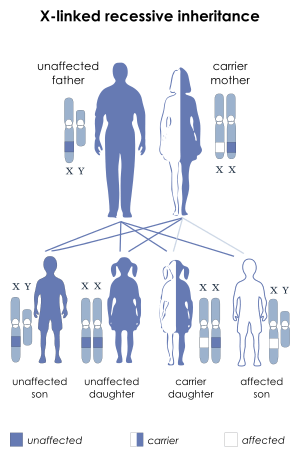
Protanopia, deuteranopia, protanomaly, and deuteranomaly are commonly inherited forms of red–green color blindness which affect a substantial portion of the human population. Those affected have difficulty with discriminating red and green hues due to the absence or mutation of the red or green retinal photoreceptors. This deficiency does not cause difficulty discerning red from green. [11][20] It is sex-linked: genetic red–green color blindness affects males much more often than females, because the genes for the red and green color receptors are located on the X chromosome, of which males have only one and females have two. Females (46, XX) are red–green color blind only if both their X chromosomes are defective with a similar deficiency, whereas males (46, XY) are color blind if their single X chromosome is defective.
The gene for red–green color blindness is transmitted from a color blind male to all his daughters who are heterozygote carriers and are usually unaffected. In turn, a carrier woman has a fifty percent chance of passing on a mutated X chromosome region to each of her male offspring. The sons of an affected male will not inherit the trait from him, since they receive his Y chromosome and not his (defective) X chromosome. Should an affected male have children with a carrier or colorblind woman, their daughters may be colorblind by inheriting an affected X chromosome from each parent.
Because one X chromosome is inactivated at random in each cell during a woman's development, it is possible for her to have four different cone types, as when a carrier of protanomaly has a child with a deuteranomalic man. Denoting the normal vision alleles by P and D and the anomalous by p and d, the carrier is PD pD and the man is Pd. The daughter is either PD Pd or pD Pd. Suppose she is pD Pd. Each cell in her body expresses either her mother's chromosome pD or her father's Pd. Thus her red–green sensing will involve both the normal and the anomalous pigments for both colors. Such females are tetrachromats, since they require a mixture of four spectral lights to match an arbitrary light.
Red–green color blindness can be caused by ethambutol.[21]
- Protanopia (1% of males): Lacking the red cones for long-wavelength sensitive retinal cones, those with this condition are unable to distinguish between colors in the green–yellow–red section of the spectrum. They have a neutral point at a cyan-like wavelength around 492 nm (see spectral color for comparison) – that is, they cannot discriminate light of this wavelength from white. For a protanope, the brightness of red, orange, and yellow are much reduced compared to normal. This dimming can be so pronounced that reds may be confused with black or dark gray, and red traffic lights may appear to be extinguished. They may learn to distinguish reds from yellows primarily on the basis of their apparent brightness or lightness, not on any perceptible hue difference. Violet, lavender, and purple are indistinguishable from various shades of blue because their reddish components are so dimmed as to be invisible. For example, pink flowers, reflecting both red light and blue light, may appear just blue to the protanope. A very few people have been found who have one normal eye and one protanopic eye. These unilateral dichromats report that with only their protanopic eye open, they see wavelengths shorter than neutral point as blue and those longer than it as yellow. This is a rare form of color blindness.
- Deuteranopia (1% of males): Lacking the green cones for medium-wavelength cones, those affected are again unable to distinguish between colors in the green–yellow–red section of the spectrum. Their neutral point is at a slightly longer wavelength, 498 nm, a more greenish hue of cyan. A deuteranope suffers the same hue discrimination problems as protanopes, but without the abnormal dimming. Purple colors are not perceived as something opposite to spectral colors; all these appear similarly. This form of colorblindness is also known as Daltonism after John Dalton (his diagnosis was confirmed as deuteranopia in 1995, some 150 years after his death, by DNA analysis of his preserved eyeball). Equivalent forms for daltonism in Romanic languages such as daltonismo (Spanish, Portuguese and Italian), daltonisme (French), daltonism (Romanian) are still used to describe color blindess in a broad sense or deuteranopia in a more restricted sense. Deuteranopic unilateral dichromats report that with only their deuteranopic eye open, they see wavelengths shorter than neutral point as blue and longer than it as yellow.[22]
- Protanomaly (1% of males, 0.01% of females):[23] Having a mutated form of the long-wavelength (red) pigment, whose peak sensitivity is at a shorter wavelength than in the normal retina, protanomalous individuals are less sensitive to red light than normal. This means that they are less able to discriminate colors, and they do not see mixed lights as having the same colors as normal observers. They also suffer from a darkening of the red end of the spectrum. This causes reds to reduce in intensity to the point where they can be mistaken for black. Protanomaly is a fairly rare form of color blindness, making up about 1% of the male population. Both protanomaly and deuteranomaly are carried on the X chromosome.
- Deuteranomaly (most common—6% of males, 0.4% of females):[23] These individuals have a mutated form of the medium-wavelength (green) pigment. The medium-wavelength pigment is shifted towards the red end of the spectrum resulting in a reduction in sensitivity to the green area of the spectrum. Unlike protanomaly the intensity of colors is unchanged. The deuteranomalous person is considered "green weak". For example, in the evening, dark green cars appear to be black to Deuteranomalous people. Similar to the protanomates, deuteranomates are poor at discriminating small differences in hues in the red, orange, yellow, green region of the spectrum. They make errors in the naming of hues in this region because the hues appear somewhat shifted towards green. One very important difference between deuteranomalous individuals and protanomalous individuals is deuteranomalous individuals do not have the loss of "brightness" problem.
Blue–yellow color blindness
Those with tritanopia and tritanomaly have difficulty discriminating between bluish and greenish hues, as well as yellowish and reddish hues.
Color blindness involving the inactivation of the short-wavelength sensitive cone system (whose absorption spectrum peaks in the bluish-violet) is called tritanopia or, loosely, blue–yellow color blindness. The tritanope's neutral point occurs near a yellowish 570 nm; green is perceived at shorter wavelengths and red at longer wavelengths.[24] Mutation of the short-wavelength sensitive cones is called tritanomaly. Tritanopia is equally distributed among males and females. Jeremy H. Nathans (with the Howard Hughes Medical Institute) demonstrated that the gene coding for the blue receptor lies on chromosome 7, which is shared equally by males and females. Therefore, it is not sex-linked. This gene does not have any neighbor whose DNA sequence is similar. Blue color blindness is caused by a simple mutation in this gene.
- Tritanopia (less than 1% of males and females): Lacking the short-wavelength cones, those affected see short-wavelength colors (blue, indigo and a spectral violet) greenish and drastically dimmed, some of these colors even as black. Yellow is indistinguishable from pink, and purple colors are perceived as various shades of red. This form of color blindness is not sex-linked.
- Tritanomaly (equally rare for males and females [0.01% for both]):[23] Having a mutated form of the short-wavelength (blue) pigment. The short-wavelength pigment is shifted towards the green area of the spectrum. This is the rarest form of anomalous trichromacy color blindness. Unlike the other anomalous trichromacy color deficiencies, the mutation for this color blindness is carried on chromosome 7. Therefore, it is equally prevalent in both male & female populations. The OMIM gene code for this mutation is 304000 "Colorblindness, Partial Tritanomaly".[25]
Total color blindness
Total color blindness is defined as the inability to see color. Although the term may refer to acquired disorders such as cerebral achromatopsia also known as color agnosia, it typically refers to congenital color vision disorders (i.e. more frequently rod monochromacy and less frequently cone monochromacy).[26][27]
In cerebral achromatopsia, a person cannot perceive colors even though the eyes are capable of distinguishing them. Some sources do not consider these to be true color blindness, because the failure is of perception, not of vision. They are forms of visual agnosia.[27]
Monochromacy is the condition of possessing only a single channel for conveying information about color. Monochromats possess a complete inability to distinguish any colors and perceive only variations in brightness. It occurs in two primary forms:
- Rod monochromacy, frequently called achromatopsia, where the retina contains no cone cells, so that in addition to the absence of color discrimination, vision in lights of normal intensity is difficult. While normally rare, achromatopsia is very common on the island of Pingelap, a part of the Pohnpei state, Federated States of Micronesia, where it is called maskun: about 10% of the population there has it, and 30% are unaffected carriers. The island was devastated by a storm in the 18th century (an example of a genetic bottleneck) and one of the few male survivors carried a gene for achromatopsia. The population grew to several thousand before foreign troops introduced diseases to the island in the 1940s.
- Cone monochromacy is the condition of having both rods and cones, but only a single kind of cone. A cone monochromat can have good pattern vision at normal daylight levels, but will not be able to distinguish hues. Blue cone monochromacy (X chromosome) is caused by lack of functionality of L and M cones (red and green). It is encoded at the same place as red–green color blindness on the X chromosome. Peak spectral sensitivities are in the blue region of the visible spectrum (near 440 nm). People with this condition generally show nystagmus ("jiggling eyes"), photophobia (light sensitivity), reduced visual acuity, and myopia (nearsightedness).[28] Visual acuity usually falls to the 20/50 to 20/400 range.
Mechanism
The typical human retina contains two kinds of light cells: the rod cells (active in low light) and the cone cells (active in normal daylight). Normally, there are three kinds of cone cells, each containing a different pigment, which are activated when the pigments absorb light. The spectral sensitivities of the cones differ; one is most sensitive to short wavelengths, one to medium wavelengths, and the third to medium-to-long wavelengths within the visible spectrum, with their peak sensitivities in the blue, green, and yellow-green regions of the spectrum, respectively. The absorption spectra of the three systems overlap, and combine to cover the visible spectrum. These receptors are known as short (S), medium (M),and long (L) wavelength cones, but are also often referred to as blue, green, and red cones, although this terminology is inaccurate.[29]
The receptors are each responsive to a wide range of wavelengths. For example, the long wavelength "red" receptor has its peak sensitivity in the yellow-green, some way from the red end (longest wavelength) of the visible spectrum. The sensitivity of normal color vision actually depends on the overlap between the absorption ranges of the three systems: different colors are recognized when the different types of cone are stimulated to different degrees. Red light, for example, stimulates the long wavelength cones much more than either of the others, and reducing the wavelength causes the other two cone systems to be increasingly stimulated, causing a gradual change in hue.
Many of the genes involved in color vision are on the X chromosome, making color blindness much more common in males than in females because males only have one X chromosome, while females have two. Because this is an X-linked trait, an estimated 2–3% of women have a 4th color cone[30] and can be considered tetrachromats. One such woman has been reported to be a true or functional tetrachromat, as she can discriminate colors most other people can't.[31][32]
Diagnosis
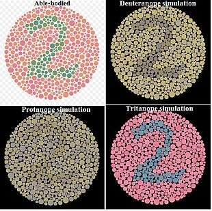
The Ishihara color test, which consists of a series of pictures of colored spots, is the test most often used to diagnose red–green color deficiencies.[33] A figure (usually one or more Arabic digits) is embedded in the picture as a number of spots in a slightly different color, and can be seen with normal color vision, but not with a particular color defect. The full set of tests has a variety of figure/background color combinations, and enable diagnosis of which particular visual defect is present. The anomaloscope, described above, is also used in diagnosing anomalous trichromacy.
Position yourself about 75cm from your monitor so that the colour test image you are looking at is at eye level, read the description of the image and see what you can see!! It is not necessary in all cases to use the entire set of images. In a large scale examination the test can be simplified to 6 tests; test, one of tests 2 or 3, one of tests 4, 5, 6 or 7, one of tests 8 or 9, one of tests 10, 11, 12 or 13 and one of tests 14 or 15.
Because the Ishihara color test contains only numerals, it may not be useful in diagnosing young children, who have not yet learned to use numerals. In the interest of identifying these problems early on in life, alternative color vision tests were developed using only symbols (square, circle, car).
Besides the Ishihara color test, the US Navy and US Army also allow testing with the Farnsworth Lantern Test. This test allows 30% of color deficient individuals, whose deficiency is not too severe, to pass.
Another test used by clinicians to measure chromatic discrimination is the Farnsworth-Munsell 100 hue test. The patient is asked to arrange a set of colored caps or chips to form a gradual transition of color between two anchor caps.[34]
The HRR color test (developed by Hardy, Rand, and Rittler) is a red–green color test that, unlike the Ishihara, also has plates for the detection of the tritan defects.[35]
Most clinical tests are designed to be fast, simple, and effective at identifying broad categories of color blindness. In academic studies of color blindness, on the other hand, there is more interest in developing flexible tests to collect thorough datasets, identify copunctal points, and measure just noticeable differences.[36]
Management
There is generally no treatment to cure color deficiencies. ″The American Optometric Association reports a contact lens on one eye can increase the ability to differentiate between colors, though nothing can make you truly see the deficient color.″[37]
Lenses
Optometrists can supply colored spectacle lenses or a single red-tint contact lens to wear on the non-dominant eye, but although this may improve discrimination of some colors, it can make other colors more difficult to distinguish. A 1981 review of various studies to evaluate the effect of the X-chrom contact lens concluded that, while the lens may allow the wearer to achieve a better score on certain color vision tests, it did not correct color vision in the natural environment.[38] A case history using the X-Chrom lens for a rod monochromat is reported[39] and an X-Chrom manual is online.[40]
Lenses that filter certain wavelengths of light can allow people with a cone anomaly, but not dichromacy, to see a better spectrum of colors, especially those with classic "red/green" color blindness. They work by notching out wavelengths that strongly stimulate both red and green cones in a deuter- or protanomalous person, improving the distinction between the two cones' signals. As of 2013, sunglasses that enhance colors for many colorblind people are available commercially.[41][42][43]
Apps
Many applications for iPhone and iPad have been developed to help colorblind people to view the colors in a better way. Many applications launch a sort of simulation of colorblind vision to make normal-view people understand how the color-blinds see the world. Others allow a correction of the image grabbed from the camera with a special "daltonizer" algorithm.
The GNOME desktop environment provides colorblind accessibility using the gnome-mag and the libcolorblind software. Using a gnome applet, the user may switch a color filter on and off, choosing from a set of possible color transformations that will displace the colors in order to disambiguate them. The software enables, for instance, a colorblind person to see the numbers in the Ishihara test.
Epidemiology
| Males | Females | |
|---|---|---|
| Dichromacy | 2.4% | 0.03% |
| Protanopia (red deficient: L cone absent) | 1.3% | 0.02% |
| Deuteranopia (green deficient: M cone absent) | 1.2% | 0.01% |
| Tritanopia (blue deficient: S cone absent) | 0.001% | 0.03% |
| Anomalous trichromacy | 6.3% | 0.37% |
| Protanomaly (red deficient: L cone defect) | 1.3% | 0.02% |
| Deuteranomaly (green deficient: M cone defect) | 5.0% | 0.35% |
| Tritanomaly (blue deficient: S cone defect) | 0.0001% | 0.0001% |
Color blindness affects a large number of individuals, with protanopia and deuteranopia being the most common types.[11] In individuals with Northern European ancestry, as many as 8 percent of men and 0.4 percent of women experience congenital color deficiency.[45]
The number affected varies among groups. Isolated communities with a restricted gene pool sometimes produce high proportions of color blindness, including the less usual types. Examples include rural Finland, Hungary, and some of the Scottish islands. In the United States, about 7 percent of the male population—or about 10.5 million men—and 0.4 percent of the female population either cannot distinguish red from green, or see red and green differently from how others do (Howard Hughes Medical Institute, 2006 ). More than 95 percent of all variations in human color vision involve the red and green receptors in male eyes. It is very rare for males or females to be "blind" to the blue end of the spectrum.[46]
Red–green color blindness
| Population | N | % |
| Arabs (Druzes) | 337 | 10.0 |
| Aboriginal Australians | 4,455 | 1.9 |
| Belgians | 9,540 | 7.4 |
| Bosnians | 4,836 | 6.2 |
| Britons | 16,180 | 6.6 |
| Chinese | 1,164 | 6.9 |
| Dutch | 3,168 | 8.0 |
| Eskimo | 297 | 2.5 |
| Fiji Islanders | 608 | 0.8 |
| French | 1,243 | 8.6 |
| Germans | 7,861 | 7.7 |
| Hutu | 1,000 | 2.9 |
| Indians (Andhra Pradesh) | 292 | 7.5 |
| Iranians | 16,180 | 6.6 |
| Japanese | 259,000 | 4.0 |
| Navajo | 571 | 2.3 |
| Norwegians | 9,047 | 9.0 |
| Mexicans | 571 | 2.3 |
| Russians | 1,343 | 9.2 |
| Scots | 463 | 7.8 |
| Swiss | 2,000 | 8.0 |
| Tibetans | 241 | 5.0 |
| Tswana | 407 | 2.0 |
| Tutsi | 1,000 | 2.5 |
| Vojvodinians | 4,750 | 7.4 |
| DR Congolese | 929 | 1.7 |
History

The first scientific paper on the subject of color blindness, Extraordinary facts relating to the vision of colours, was published by the English chemist John Dalton in 1798[49] after the realization of his own color blindness. Because of Dalton's work, the general condition has been called daltonism, although in English this term is now used only for deuteranopia.
Society and culture
Design implications
Color codes present particular problems for those with color deficiencies as they are often difficult or impossible for them to perceive.
Good graphic design avoids using color coding or using color contrasts alone to express information;[50] not only helps color blind people, but also aids understanding by normally sighted people.
Designers need to take into account that color-blindness is highly sensitive to differences in material. For example, a red–green colorblind person who is incapable of distinguishing colors on a map printed on paper may have no such difficulty when viewing the map on a computer screen or television. In addition, some color blind people find it easier to distinguish problem colors on artificial materials, such as plastic or in acrylic paints, than on natural materials, such as paper or wood. Third, for some color blind people, color can only be distinguished if there is a sufficient "mass" of color: thin lines might appear black, while a thicker line of the same color can be perceived as having color.
Designers should also note that red–blue and yellow–blue color combinations are generally safe. So instead of the ever popular "red means bad and green means good" system, using these combinations can lead to a much higher ability to use color coding effectively. This will still cause problems for those with monochromatic color blindness, but it is still something worth considering.[51]
When the need to process visual information as rapidly as possible arises, for example in an emergency situation, the visual system may operate only in shades of gray, with the extra information load in adding color being dropped. This is an important possibility to consider when designing, for example, emergency brake handles or emergency phones.
Occupations
Color blindness may make it difficult or impossible for a person to engage in certain occupations. Persons with color blindness may be legally or practically barred from occupations in which color perception is an essential part of the job (e.g., mixing paint colors), or in which color perception is important for safety (e.g., operating vehicles in response to color-coded signals). This occupational safety principle originates from the Lagerlunda train crash of 1875 in Sweden. Following the crash, Professor Alarik Frithiof Holmgren, a physiologist, investigated and concluded that the color blindness of the engineer (who had died) had caused the crash. Professor Holmgren then created the first test using different-colored skeins to exclude people from jobs in the transportation industry on the basis of color blindness.[52] However, there is a claim that there is no firm evidence that color deficiency did cause the collision, or that it might have not been the sole cause.[53]
Color vision is important for occupations using telephone or computer networking cabling, as the individual wires inside the cables are color-coded using green, orange, brown, blue and white colors.[54] Electronic wiring, transformers, resistors, and capacitors are color-coded as well, using black, brown, red, orange, yellow, green, blue, violet, gray, white, silver, gold.[55]
Driving
Some countries (for example, Romania) have refused to grant driving licenses to individuals with color blindness. In Romania, there is an ongoing campaign to remove the legal restrictions that prohibit colorblind citizens from getting drivers' licenses.[56]
The usual justification for such restrictions is that drivers of motor vehicles must be able to recognize color-coded signals, such as traffic lights or warning lights.[57]
Piloting aircraft
While many aspects of aviation depend on color coding, only a few of them are critical enough to be interfered with by some milder types of color blindness. Some examples include color-gun signaling of aircraft that have lost radio communication, color-coded glide-path indications on runways, and the like. Some jurisdictions restrict the issuance of pilot credentials to persons who suffer from color blindness for this reason. Restrictions may be partial, allowing color-blind persons to obtain certification but with restrictions, or total, in which case color-blind persons are not permitted to obtain piloting credentials at all.
In the United States, the Federal Aviation Administration requires that pilots be tested for normal color vision as part of their medical clearance in order to obtain the required medical certificate, a prerequisite to obtaining a pilot's certification. If testing reveals color blindness, the applicant may be issued a license with restrictions, such as no night flying and no flying by color signals—such a restriction effectively prevents a pilot from holding certain flying occupations, such as that of an airline pilot, although commercial pilot certification is still possible, and there are a few flying occupations that do not require night flight and thus are still available to those with restrictions due to color blindness (e.g., agricultural aviation). The government allows several types of tests, including medical standard tests (e.g., the Ishihara, Dvorine, and others) and specialized tests oriented specifically to the needs of aviation. If an applicant fails the standard tests, they will receive a restriction on their medical certificate that states: "Not valid for night flying or by color signal control". They may apply to the FAA to take a specialized test, administered by the FAA. Typically, this test is the "color vision light gun test". For this test an FAA inspector will meet the pilot at an airport with an operating control tower. The color signal light gun will be shone at the pilot from the tower, and they must identify the color. If they pass they may be issued a waiver, which states that the color vision test is no longer required during medical examinations. They will then receive a new medical certificate with the restriction removed. This was once a Statement of Demonstrated Ability (SODA), but the SODA was dropped, and converted to a simple waiver (letter) early in the 2000s.[58]
Research published in 2009 carried out by the City University of London's Applied Vision Research Centre, sponsored by the UK's Civil Aviation Authority and the US Federal Aviation Administration, has established a more accurate assessment of color deficiencies in pilot applicants' red–green and yellow–blue color range which could lead to a 35% reduction in the number of prospective pilots who fail to meet the minimum medical threshold.[59]
Art
Inability to distinguish color does not necessarily preclude the ability to become a celebrated artist. The 20th century expressionist painter Clifton Pugh, three-time winner of Australia's Archibald Prize, on biographical, gene inheritance and other grounds has been identified as a protanope.[60] 19th century French artist Charles Méryon became successful by concentrating on etching rather than painting after he was diagnosed as having a red–green deficiency.[61]
Rights of the color blind
A famous traffic light on Tipperary Hill in Syracuse, New York, is upside-down due to the sentiments of its Irish American community, but has been criticized due to the potential hazard it poses for color-blind persons.[62]
Brazil
A Brazilian court ruled that people with color blindness are protected by the Inter-American Convention on the Elimination of All Forms of Discrimination against Person with Disabilities.[63][64][65]
At trial, it was decided that the carriers of color blindness have a right of access to wider knowledge, or the full enjoyment of their human condition.
Research
Some tentative evidence finds that color blind people are better at penetrating certain color camouflages. Such findings may give an evolutionary reason for the high rate of red–green color blindness.[3] There is also a study suggesting that people with some types of color blindness can distinguish colors that people with normal color vision are not able to distinguish.[66] In World War II, color blind observers were used to penetrate camouflage.[67]
In September 2009, the journal Nature reported that researchers at the University of Washington and University of Florida were able to give trichromatic vision to squirrel monkeys, which normally have only dichromatic vision, using gene therapy.[68]
In 2003, a cybernetic device called eyeborg was developed to allow the wearer to hear sounds representing different colors.[69] Achromatopsic artist Neil Harbisson was the first to use such a device in early 2004; the eyeborg allowed him to start painting in color by memorizing the sound corresponding to each color. In 2012, at a TED Conference, Harbisson explained how he could now perceive colors outside the ability of human vision.[70] Portuguese Designer Miguel Neiva developed a code system, named Coloradd, based on five basic shapes that, when combined, make it easier to identify various colors for colorblind people. Its use is currently expanding in Portugal (hospitals, transportation, education) and in other countries.
See also
References
- 1 2 3 Gordon, N (March 1998). "Colour blindness.". Public health. 112 (2): 81–4. doi:10.1038/sj.ph.1900446. PMID 9581449.
- 1 2 3 4 5 6 7 "Facts About Color Blindness". NEI. February 2015. Retrieved 29 July 2016.
- 1 2 Morgan, M. J.; Adam, A.; Mollon, J. D. (June 1992). "Dichromats detect colour-camouflaged objects that are not detected by trichromats". Proc. Biol. Sci. 248 (1323): 291–5. doi:10.1098/rspb.1992.0074. PMID 1354367.
- ↑ Acquired Colour Vision Defects. colourblindawareness.org
- ↑ MedlinePlus Encyclopedia Color blindness
- ↑ "Types of Color Deficiencies". Konan Medical. Retrieved 2014-04-26.
- ↑ Protanopia – Red–Green Color Blindness. color-blindness.com
- ↑ Deuteranopia – Red–Green Color Blindness. color-blindness.com
- ↑ Tovee, Martin J. (2008). An Introduction to the Visual System. Cambridge University Press. ISBN 0-521-70964-4.
- ↑ Tritanopia – Blue–Yellow Color Blindness. color-blindness.com
- 1 2 3 Wong, Bang (2011). "Color blindness". Nature Methods. 8 (6): 441. doi:10.1038/nmeth.1618. PMID 21774112.
- ↑ Albrecht, Mario (2010). "Color blindness". Nature Methods. 7 (10): 775–775. doi:10.1038/nmeth1010-775a. ISSN 1548-7091.
- ↑ Sharpe, L.T.; Stockman, A.; Jägle, H.; Nathans, J. (1999). "Opsin genes, cone photopigments, color vision and color blindness". In Gegenfurtner, K. R.; Sharpe, L. T. Color Vision: From Genes to Perception. Cambridge University Press. ISBN 978-0-521-00439-8.
- ↑ American Medical Association (2003). Leikin, Jerrold B.; Lipsky, Martin S., eds. Complete Medical Encyclopedia (encyclopedia) (First ed.). New York, NY: Random House Reference. p. 388. ISBN 0-8129-9100-1.
- ↑ Spring, Kenneth R.; Parry-Hill, Matthew J.; Fellers, Thomas J.; Davidson, Michael W. "Human Vision and Color Perception". Florida State University. Retrieved 2007-04-05.
- ↑ Hoffman, Paul S. "Accommodating Color Blindness" (PDF). Archived from the original (PDF) on 15 May 2008. Retrieved 2009-07-01.
- ↑ Neitz, Maureen E. "Severity of Colorblindness Varies". Medical College of Wisconsin. Archived from the original on 5 February 2007. Retrieved 2007-04-05.
- ↑ Jones, Sara A; Shim, Sang-Hee; He, Jiang; Zhuang, Xiaowei (2011). "Fast, three-dimensional super-resolution imaging of live cells". Nature Methods. 8 (6): 499–508. doi:10.1038/nmeth.1605. PMC 3137767
 . PMID 21552254.
 - ↑ Simunovic, M P (2010). "Colour vision deficiency". Eye. 24 (5): 747–55. doi:10.1038/eye.2009.251. PMID 19927164.
- ↑ Neitz, Jay; Neitz, Maureen (2011). "The genetics of normal and defective color vision". Vision Research. 51 (7): 633–51. doi:10.1016/j.visres.2010.12.002. PMC 3075382. PMID 21167193.
- ↑ "Myambutol (Ethambutol) Drug Information: Description, User Reviews, Drug Side Effects, Interactions – Prescribing Information at RxList". Rxlist.com. Retrieved 2014-05-24.
- ↑ David L. MacAdam (ed.) and Deane B. Judd (1979). Contributions to color science. NBS. p. 584.
- 1 2 3 Kalloniatis, Michael; Luu, Charles (July 9, 2007). "The Perception of Color". In Kolb, Helga; Fernandez, Eduardo; Nelson, Ralph. Webvision: The Organization of the Retina and Visual System. PMID 21413396.
- ↑ Goldstein, E. Bruce (2007). Sensation and perception (7th ed.). Wadsworth: Thomson. p. 152. ISBN 978-0-534-55810-9.
- ↑ "Disease-causing Mutations and protein structure". UCL Biochemistry BSM Group. Retrieved 2007-04-02.
- ↑ "Types of Colour Blindness". Colour Blind Awareness.
- 1 2 Blom, Jan Dirk (2009). A Dictionary of Hallucinations. Springer. p. 4. ISBN 978-1-4419-1222-0.
- ↑ Weiss, A. H.; Biersdorf, W. R. (1989). "Blue cone monochromatism". J Pediatr Ophthalmol Strabismus. 26 (5): 218–23. PMID 2795409.
- ↑ "Colour vision deficiency – Causes". NHS Choices. 2012-12-14. Retrieved 2014-05-24.
- ↑ Roth, Mark (13 September 2006). "Some women may see 100,000,000 colors, thanks to their genes". Pittsburgh Post-Gazette.
- ↑ Didymus, JohnThomas (Jun 19, 2012), "Scientists find woman who sees 99 million more colors than others", Digital Journal
- ↑ Jordan, Gabriele; Deeb, Samir S.; Bosten, Jenny M.; Mollon, J. D. (July 2010). "The dimensionality of color vision in carriers of anomalous trichromacy". Journal of Vision. 10 (12): 12. doi:10.1167/10.8.12. PMID 20884587.
- ↑ Gordon, N (1998). "Colour blindness". Public Health. 112 (2): 81–4. doi:10.1038/sj.ph.1900446. PMID 9581449.
- ↑ Kinnear, PR; Sahraie, A (2002). "New Farnsworth-Munsell 100 hue test norms of normal observers for each year of age 5–22 and for age decades 30–70". The British Journal of Ophthalmology. 86 (12): 1408–11. doi:10.1136/bjo.86.12.1408. PMC 1771429. PMID 12446376.
- ↑ Cole, Barry L; Lian, Ka-Yee; Lakkis, Carol (2006). "The new Richmond HRR pseudoisochromatic test for colour vision is better than the Ishihara test". Clinical and Experimental Optometry. 89 (2): 73–80. doi:10.1111/j.1444-0938.2006.00015.x. PMID 16494609.
- ↑ Toufeeq, A (2004). "Specifying colours for colour vision testing using computer graphics". Eye. 18 (10): 1001–5. doi:10.1038/sj.eye.6701378. PMID 15192692.
- ↑ Color Vision Deficiency. American Optometric Association
- ↑ Siegel, I. M. (1981). "The X-Chrom lens. On seeing red". Surv Ophthalmol. 25 (5): 312–24. PMID 6971497.
- ↑ Zeltzer, HI (1979). "Use of modified X-Chrom for relief of light dazzlement and color blindness of a rod monochromat". Journal of the American Optometric Association. 50 (7): 813–8. PMID 315420.
- ↑ An X-Chrom manual
- ↑ A Scientist Accidentally Developed Sunglasses That Could Correct Color Blindness
- ↑ Introducing EnChroma
- ↑ Pogue, David (15 August 2013). "Glasses That Solve Colorblindness, for a Big Price Tag". The New York Times. Retrieved 22 July 2015.
- ↑ "Causes and Incidence of Colorblindness". Causes of Color. Retrieved 27 February 2014.
- ↑ Chan, Xin; Goh, Shi; Tan, Ngiap (2014). "Subjects with colour vision deficiency in the community: what do primary care physicians need to know?". Asia Pacific Family Medicine. 13 (1): 10. doi:10.1186/s12930-014-0010-3.
- ↑ Ananya, Mandal. "Color Blindness Prevalence". Health. Retrieved 27 February 2014.
- ↑ Harrison et al. (1977): Human Biology, Oxford University Press, Oxford, ISBN 0-19-857164-X; ISBN 0-19-857165-8.
- ↑ Hadžiselimović R., Berberović Lj., Sofradžija A. (1980): Populacijska genetika viđenja crvenog i zelenog dijela spektra u stanovništvu Bosne i Hercegovine / Population genetics of red and green spectrum vision of the population of Bosnia and Herzegovina. God. Biol. inst. Univ. u Sarajevu / Annual of Institute of Biology, University of Sarajevo, 33: 87–97.
- ↑ Dalton, J (1798). "Extraordinary facts relating to the vision of colours: with observations". Memoirs of the Literary and Philosophical Society of Manchester. 5: 28–45. OCLC 9879327.
- ↑ Crow, Kevin L. (2008). "Four Types of Disabilities: Their Impact on Online Learning". TechTrends. 52 (1): 51–5. doi:10.1007/s11528-008-0112-6.
- ↑ Habibzadeh, Parham (2015-01-01). "Our red–green world". Australian Health Review. doi:10.1071/ah15161.
- ↑ Algis, J.; Vingrys, J.; Cole, Barry L. (1986). "Origins of colour vision standards within the transport industry". Ophthalmic & Physiological Optics. 6 (4): 369–75. doi:10.1111/j.1475-1313.1986.tb01155.x. PMID 3306566.
- ↑ Mollon, JD; Cavonius, LR (2012). "The Lagerlunda Collision and the Introduction of Color Vision Testing". Survey of Ophthalmology. 57 (2): 178–94. doi:10.1016/j.survophthal.2011.10.003. PMID 22301271.
- ↑ Meyers, Michael (2002). All in One A+ Certification Exam Guide (4th ed.). Berkeley, California: McGraw-Hill/Osborne. ISBN 0-07-222274-3.
- ↑ Grob, Bernard (2001). Basic Electronics. Columbus, Ohio: Glencoe/McGraw-Hill. ISBN 0-02-802253-X.
- ↑ "Petition to European Union on Colorblind's condition in Romania". Retrieved 2007-08-21.
- ↑ Habibzadeh, Parham (2015-01-01). "Our red–green world". Australian Health Review. doi:10.1071/ah15161.
- ↑ "Aerospace Medical Dispositions — Color vision". Retrieved 2009-04-11.
- ↑ Warburton, Simon (29 May 2009). "Colour-blindness research could clear more pilots to fly: UK CAA". Air transport. Reed Business Information. Retrieved 29 October 2009.
- ↑ Cole, Barry L; Harris, Ross W (2009). "Colour blindness does not preclude fame as an artist: celebrated Australian artist Clifton Pugh was a protanope". Clinical and Experimental Optometry. 92 (5): 421–8. doi:10.1111/j.1444-0938.2009.00384.x. PMID 19515095.
- ↑ Anon. "Charles Meryon". Art Encyclopedia. The Concise Grove Dictionary of Art. Oxford University Press. Retrieved 7 January 2010.
- ↑ Sarah Zhang. "The Story Behind Syracuse's Upside-Down Traffic Light". Gizmodo.
- ↑ "Full text of the decision of the court – in Portuguese language". Retrieved 2012-03-09.
- ↑ "Decree issued by president of a republic ratifying Legislative Decree No. 198, of june 13, which approved the Inter-American Convention AG/RES. 1608 – in Portuguese language". Retrieved 2012-03-09.
- ↑ "Inter-American Convention on the Elimination of All Forms of Discrimination against Person with Disabilities.". Retrieved 2012-03-09.
- ↑ Bosten, J.M.; Robinson, J.D.; Jordan, G.; Mollon, J.D. (2005). "Multidimensional scaling reveals a color dimension unique to 'color-deficient' observers". Current Biology. 15 (23): R950–2. doi:10.1016/j.cub.2005.11.031. PMID 16332521.
- ↑ "Colour blindness not all it seems". BBC News. 6 December 2015. Retrieved 21 June 2016.
- ↑ Dolgin, Elie (2009). "Colour blindness corrected by gene therapy". Nature. doi:10.1038/news.2009.921.
- ↑ Alfredo M. Ronchi: Eculture: Cultural Content in the Digital Age. Springer (New York, 2009). p. 319 ISBN 978-3-540-75273-8
- ↑ "I listen to color", Neil Harbisson at TED Global, 27 June 2012.
Further reading
- Kaiser, Peter K.; Boynton, Robert M. (1996). Human color vision. Washington, DC: Optical Society of America. ISBN 1-55752-461-0. OCLC 472932250.
- McIntyre, Donald (2002). Colour blindness: causes and effects. Chester: Dalton Publishing. ISBN 0-9541886-0-8. OCLC 49204679.
- Rubin, Melvin L.; Cassin, Barbara; Solomon, Sheila (1984). Dictionary of eye terminology. Gainesville, Fla: Triad Pub. Co. ISBN 0-937404-07-1. OCLC 10375427.
- Shevell, Steven K. (2003). The science of color. Amsterdam: Elsevier. ISBN 0-444-51251-9. OCLC 52271315.
- Hilbert, David; Byrne, Alexander (1997). Readings on color. Cambridge, Mass: MIT Press. ISBN 0-262-52231-4. OCLC 35762680.
- Stiles, W. S.; Wyszecki, Günter (2000). Color science: concepts and methods, quantitative data and formulae. Chichester: John Wiley & Sons. ISBN 0-471-39918-3. OCLC 799532137.
- Kuchenbecker, J.; Broschmann, D. (2014). Plates for color vision testing. New York: Thieme. ISBN 978-3-13-175481-3.
External links
| Wikimedia Commons has media related to Color blindness. |
| Wikisource has original text related to this article: |
Color topics | |||||||||||
|---|---|---|---|---|---|---|---|---|---|---|---|
| Color science |
| ||||||||||
| Color philosophy | |||||||||||
| Color terms |
| ||||||||||
| Color organizations | |||||||||||
| Lists | |||||||||||
| Related | |||||||||||
| |||||||||||