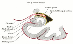
Choroid plexus
| Choroid plexus | |
|---|---|
 Scheme of roof of fourth ventricle. The arrow is in the foramen of Magendie. 1: inferior medullary velum 2: Choroid plexus 3: Cerebellomedullary cistern of subarachnoid cavity 4: Central canal 5: Corpora quadrigemina 6: Cerebral peduncle 7: Superior medullary velum 8: Ependymal lining of ventricle 9: Pontine cistern of subarachnoid cavity | |
 Coronal section of lateral and third ventricles. | |
| Details | |
| Identifiers | |
| Latin | Plexus choroideus |
| MeSH | A08.186.211.276.298 |
| NeuroNames | ancil-456 |
| NeuroLex ID | Choroid plexus |
| TA |
A14.1.09.279 A14.1.01.307 A14.1.01.306 A14.1.01.304 |
| FMA | 61934 |
The choroid plexus is a plexus of cells that produces the cerebrospinal fluid in the ventricles of the brain. The choroid plexus consists of modified ependymal cells.
Structure
There are four choroid plexuses (CP) in the brain, one in each of the ventricles. The CP consists of a layer of cuboidal epithelial cells surrounding a core of capillaries and loose connective tissue. The CP epithelial layer is continuous with the ependymal cell layer that lines the ventricles, but unlike the ependyma, the epithelial layer has tight gap junctions between the cells on the side facing the ventricle (apical surface). These gap junctions prevent the majority of substances from crossing the cell layer into the CSF; thus the CP acts as a blood–CSF barrier. The CP folds into many villi around each capillary, creating frond-like processes that project into the ventricles. The villi, along with a brush border of microvilli, greatly increases the surface area of the CP. CSF is formed as plasma is filtered from the blood through the epithelial cells. CP epithelial cells actively transport sodium, chloride and bicarbonate ions into the ventricles and water follows the resulting osmotic gradient.
Choroid plexus is present in all components of the ventricular system except for the cerebral aqueduct, frontal horn of the lateral ventricle,[1] and occipital horn of the lateral ventricle.[1]

Choroid plexus is found in the superior part of the inferior horn of the lateral ventricles. It follows up along this boundary, continuous with the inferior of the body of the lateral ventricles. It passes into the interventricular foramen, and is present at the top of the third ventricle.
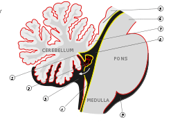
There is also choroid plexus in the fourth ventricle, in the section closest to the bottom half of the cerebellum.
The choroid plexus consists of many capillaries, separated from the ventricles by choroid epithelial cells. Fluid filters through these cells from blood to become cerebrospinal fluid. There is also much active transport of substances into, and out of, the CSF as it is made.
Function
In addition to CSF production, the CP act as a filtration system, removing metabolic waste, foreign substances, and excess neurotransmitters from the CSF. In this way the CP has a very important role in helping to maintain the delicate extracellular environment required by the brain to function optimally.
The blood–cerebrospinal fluid barrier (BCSFB)is a pair of barriers that separates peripheral and cerebral blood flow from the cerebrospinal fluid (CSF);[2] it is composed of epithelial cells of the choroid plexus at the peripheral blood–CSF boundary and the arachnoid membrane at the cerebral blood–CSF boundary.[2] The blood–CSF barrier serves the same purpose as the blood–brain barrier, but facilitates the transport of different substances into the brain due to the distinct structural characteristics between the two barrier systems.[2]
Clinical significance
Choroid plexus cysts and tumors
During embryological development, some fetuses may form choroid plexus cysts. These fluid-filled cysts can be detected by a level II ultrasound (18–20 weeks gestation). The finding is relatively common, with a prevalence of ~1%. Choroid plexus cysts (CPC) can be an isolated finding, which confers a 1% (variable based on population studied) risk of fetal aneuploidy. The risk of aneuploidy increases to 10.5-12% if other risk factors or ultrasound findings are noted. The particular size, bilaterality, disappearance/progression or the position of the CPC, do not have any effect on the risk of aneuploidy. 44-50% of Edwards syndrome (trisomy 18) cases will present with CPC, and 1.4% of Down syndrome (trisomy 21) cases will present with CPC. ~75% of abnormal karyotypes (obtained by chorionic villus sampling or amniocentesis) associated with CPCs are trisomy 18, while the remainder are trisomy 21.[3]
CPCs typically disappear later during pregnancy, and are considered soft markers. They are usually harmless, and studies have shown that they have no effect on infant and early childhood development.[4]
There are three graded types of choroid plexus tumor that mainly affect young children. These malignant neoplasms are rare.
Etymology
Choroid plexus translates from the Latin plexus chorioides,[5] which mirrors Ancient Greek χοριοειδές πλέγμα.[6] The word chorion was used by Galen to refer to the outer membrane enclosing the fetus. Both meanings of the word plexus are given as pleating, or braiding.[6] As often happens language changes and the use of both choroid or chorioid is both accepted. Nomina Anatomica (now Terminologia Anatomica) reflected this dual usage.
Additional images
-

Velum interpositum.
-

Coronal section of brain immediately in front of pons.
-

Coronal section of brain through intermediate mass of third ventricle.
-

Median sagittal section of brain.
-
Posterior and inferior cornua of left lateral ventricle exposed from the side.
-
Coronal section of inferior horn of lateral ventricle.
-

Choroid Plexus Histology 40x
-

Choroid plexus
-

Choroid plexus
See also
References
- 1 2 Young, Paul A. (2007). Basic clinical neuroscience (2nd ed.). Philadelphia, Pa.: Lippincott Williams & Wilkins. p. 292. ISBN 0-7817-5319-8.
- 1 2 3 Laterra J, Keep R, Betz LA, et al. (1999). "Blood–Cerebrospinal Fluid Barrier". Basic Neurochemistry: Molecular, Cellular and Medical Aspects. (6th ed.). Philadelphia: Lippincott-Raven.
- ↑ Drugan et al., 2000, American Journal of Medical Genetics
- ↑ Digiovanni et al., 1997, Obstetrics and Gynecology
- ↑ Suzuki, S., Katsumata, T., Ura, R. Fujita, T., Niizima, M. & Suzuki, H. (1936). Über die Nomina Anatomica Nova. Folia Anatomica Japonica, 14, 507-536.
- 1 2 Liddell, H.G. & Scott, R. (1940). A Greek-English Lexicon. revised and augmented throughout by Sir Henry Stuart Jones. with the assistance of. Roderick McKenzie. Oxford: Clarendon Press.
Sources
- Brodbelt, A; Stoodley, M (Oct 2007). "CSF pathways: a review.". British journal of neurosurgery. 21 (5): 510–20. doi:10.1080/02688690701447420. PMID 17922324.
- Strazielle, N; Ghersi-Egea, JF (Jul 2000). "Choroid plexus in the central nervous system: biology and physiopathology.". Journal of neuropathology and experimental neurology. 59 (7): 561–74. PMID 10901227.
External links
| Wikimedia Commons has media related to Choroid plexus. |
- 3-Dimensional images of choroid plexus (marked red)
- Anatomy diagram: 13048.000-3 at Roche Lexicon - illustrated navigator, Elsevier
- MedPix Images of Choroid Plexus
- More info at BrainInfo