Cervical lymph nodes
| Cervical lymph nodes | |
|---|---|
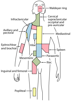 Regional lymph tissue. (Cervical near top, in green.)⋅ | |
 Deep Lymph Nodes 1. Submental 2. Submandibular (Submaxillary) Anterior Cervical Lymph Nodes (Deep) 3. Prelaryngeal 4. Thyroid 5. Pretracheal 6. Paratracheal Deep Cervical Lymph Nodes 7. Lateral jugular 8. Anterior jugular 9. Jugulodigastric Inferior Deep Cervical Lymph Nodes 10. Juguloomohyoid 11. Supraclavicular (scalene) | |
| Details | |
| Latin | Nodi lymphoidei cervicales |
Cervical lymph nodes are lymph nodes found in the neck. Of the 800 lymph nodes in the human body, 300 are in the neck.[1] Cervical lymph nodes are subject to a number of different pathological conditions including tumours, infection and inflammation.[2]
Structure
There are approximately 300 lymph nodes in the neck, and they can be classified in many different ways.[1]
Commonly used systems have been devised by the American Academy of Otolaryngology and the American Joint Committee on Cancer.[3]
One system divides the nodes as follows:[4][5]
- Level I: Submental and submandibular nodes
- Level Ia: Submental - found between the anterior belly of the digastric muscles
- Level Ib: Submandibular triangle - found around submandibular glands in submandibular space
- Level II: Upper jugular nodes - Between posterior belly of digastric muscles superiorly and hyoid bone inferiorly
- Level IIa: Anterior, medial, lateral or posterior to internal jugular vein, or if posterior, must not have an intervening fat plane
- Level IIb: Posterior to internal jugular vein with fat plane between nodes and internal jugular vein
- Level III: Middle jugular nodes - between the hyoid bone and cricoid cartilage
- Level IV: Lower jugular nodes - between the cricoid cartilage and the clavicle
- Level V: Posterior cervical or spinal accessory nodes, posterior to the sternocleidomastoid muscle
- Level VA: Spinal accessory nodes from skull base to bottom of cricoid cartilage
- Level VB: Spinal accessory nodes between cricoid and clavicle
- Level VI: Visceral space lymph nodes - midline group of cervical nodes from hyoid to sternal manubrium, includes prelaryngeal, pretracheal, and paratracheal subgroups
- Level VII: Superior mediastinal nodes - between carotid arteries from top of manubrium superiorly to innominate vein inferiorly
Clinical significance
Infectious mononucleosis (glandular fever) affects the cervical lymph nodes which become swollen.
The characterization of cancerous lymph nodes on CT scan, MRI or ultrasound is difficult, and usually requires confirmation by other nuclear imaging techniques such as PET scans. Tissue diagnosis by fine needle aspiration (which has a high rate of accuracy), may also be required. Cervical lymph node metastasis is a common feature of papillary thyroid carcinoma[6][7]
History
Henri Rouvière produced an influential classification in 1938.[8] However, this system was based upon anatomical landmarks found in dissection, making it imperfectly suited to the needs of clinicians, which led to new terminology for the lymph nodes that could be palpated.
More recently, classification systems have been proposed organized around what can be observed via diagnostic imaging.[9]
Additional images
-
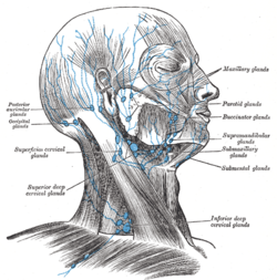
Superficial lymph glands and lymphatic vessels of head and neck.
-

Lymphatics of pharynx.
-

The lymphatics of the face.
References
- 1 2 "I. Classification". Retrieved 2008-02-16.
- ↑ Eisenmenger, LB; Wiggins RH, 3rd (January 2015). "Imaging of head and neck lymph nodes.". Radiologic clinics of North America. 53 (1): 115–32. doi:10.1016/j.rcl.2014.09.011. PMID 25476176.
- ↑ Som PM, Curtin HD, Mancuso AA (1999). "An imaging-based classification for the cervical nodes designed as an adjunct to recent clinically based nodal classifications". Arch. Otolaryngol. Head Neck Surg. 125 (4): 388–96. doi:10.1001/archotol.125.4.388. PMID 10208676.
- ↑ "Neck Dissection". Retrieved 2008-02-16.
- ↑ archotol.ama-assn.org
- ↑ Chen, C. C.; Lin, J. C.; Chen, K. W. (2015). "Lymph node ratio as a prognostic factor in head and neck cancer patients". Radiation Oncology. 10: 181. doi:10.1186/s13014-015-0490-9. PMC 4554293
 . PMID 26302761.
 - ↑ Park, C. H.; Song, C. M.; Ji, Y. B.; Pyo, J. Y.; Yi, K. J.; Song, Y. S.; Park, Y. W.; Tae, K (2015). "Significance of the Extracapsular Spread of Metastatic Lymph Nodes in Papillary Thyroid Carcinoma". Clinical and Experimental Otorhinolaryngology. 8 (3): 289–94. doi:10.3342/ceo.2015.8.3.289. PMC 4553362. PMID 26330926.
- ↑ Rouvière H. Lymphatic system of the head and neck. Tobias M, Translator. Ann Arbor, MI: Edwards Brothers, 1938.
- ↑ Chong V (2004). "Cervical lymphadenopathy: what radiologists need to know". Cancer Imaging. 4 (2): 116–20. doi:10.1102/1470-7330.2004.0020. PMC 1434593. PMID 18250018.
External links
- MedEd at Loyola medicine/pulmonar/PD/pstep23.htm
- http://www.patient.co.uk/showdoc/27000835/
- anatomy of the cervical lymphatics