Central retinal artery
| Central retinal artery | |
|---|---|
 The ophthalmic artery and its branches. (Central retinal artery visible at center.) | |
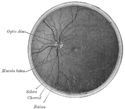 Interior of posterior half of bulb of left eye. The veins are darker in appearance than the arteries. (Central retinal artery visible but not labeled.) | |
| Details | |
| Source | ophthalmic artery |
| Vein | central retinal vein |
| Supplies | retina |
| Identifiers | |
| Latin | arteria centralis retinae |
| MeSH | A07.231.114.765 |
| TA | A12.2.06.024 |
| FMA | 49879 |
The central retinal artery (retinal artery) branches off the ophthalmic artery, running inferior to the optic nerve within its dural sheath to the eyeball.
Structure
It pierces the optic nerve close to the eyeball, sending branches over the internal surface of the retina, and these terminal branches are the only blood supply to the larger part of it.
The central part of the retina where the light rays are focussed after passing through the pupil and the lens is a circular area called the macula. The center of this circular area is the fovea. The fovea and a small area surrounding it are not supplied by the central retinal artery or its branches, but instead by the choroid.
The central retinal artery is approximately 160 micrometres in diameter.[1][2]
Variation
In some cases - approximately 20% of the population - there is a branch of the ciliary circulation called the cilio-retinal artery which supplies the retina between the macula and the optic nerve, including the nerve fibers from the foveal photoreceptors. If this artery is present, the central vision will be preserved even in case of CRAO.
Development
The central retinal artery is formed from the proximal part of the hyaloid artery after atrophy of its distal part has formed the hyaloid canal.[3]
Function
The central retinal artery supplies all the nerve fibers that form the optic nerve that carries the visual information to the lateral geniculate nucleus of the thalamus, including those that reach over the fovea.
Clinical significance
Thus if the central retinal artery gets occluded, there is complete loss of vision in that eye even though the fovea is not affected. The entire retina (with the exception of the fovea) becomes pale and swollen and opaque while the central fovea still appears reddish (this is because the choroid color shows through). This is the basis of the famous "Cherry red spot" seen on examination of the retina on funduscopy of a central retinal artery occlusion (CRAO).
However it should be remembered that the Cilio retinal artery itself is a branch of the Short Posterior Ciliary Arteries which is derived from the Ophthalmic Artery. Therefore, its possible for the cilio retinal artery itself to occlude causing significant visual loss in the perfused macula region (surrounding visual field will remain intact).
Other animals
In dogs, it is the continuation of the long ciliary artery.
Additional images
-

Horizontal section of the eyeball.
-

The terminal portion of the optic nerve and its entrance into the eyeball, in horizontal section.
-
Eye vessels
References
- ↑ Lee, K. E.; Klein, B. E. K.; Klein, R.; Meuer, S. M. (2007). "Association of Retinal Vessel Caliber to Optic Disc and Cup Diameters". Investigative Ophthalmology & Visual Science. 48 (1): 63–7. doi:10.1167/iovs.05-1203. PMID 17197517.
- ↑ Dorner, G. T.; Polska, E; Garhöfer, G; Zawinka, C; Frank, B; Schmetterer, L (2002). "Calculation of the diameter of the central retinal artery from noninvasive measurements in humans". Current eye research. 25 (6): 341–5. PMID 12789540.
- ↑ Larsen's human embryology 2009, p. 610
External links
- Diagram at suncoastretina.com
- emerg/777 at eMedicine ("Retinal artery occlusion")