Cell division
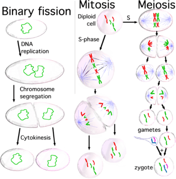
Cell division is the process by which a parent cell divides into two or more daughter cells.[1] Cell division usually occurs as part of a larger cell cycle. In eukaryotes, there are two distinct types of cell division: a vegetative division, whereby each daughter cell is genetically identical to the parent cell (mitosis),[2] and a reproductive cell division, whereby the number of chromosomes in the daughter cells is reduced by half to produce haploid gametes (meiosis). Meiosis results in twelve haploid daughter cells by undergoing one round of DNA replication followed by two divisions: homologous chromosomes are separated in the first division, and sister chromatids are separated in the second division.[3] Both of these cell division cycles are used in sexually reproducing organisms at some point in their life cycle, and both are believed to be present in the last eukaryotic common ancestor. Prokaryotes also undergo a vegetative cell division known as binary fission, where their genetic material is segregated equally into two daughter cells. All cell divisions, regardless of organism, are preceded by a single round of DNA replication.
For simple unicellular organisms[Note 1] such as the amoeba, one cell division is equivalent to reproduction – an entire new organism is created. On a larger scale, mitotic cell division can create progeny from multicellular organisms, such as plants that grow from cuttings. Mitotic cell division also enables sexually reproducing organisms to develop from the one-celled zygote, which itself was produced by meiotic cell division from gametes. After growth, cell division by mitosis allows for continual construction and repair of the organism.[4] The human body experiences about 10 quadrillion cell divisions in a lifetime.[5]
The primary concern of cell division is the maintenance of the original cell's genome. Before division can occur, the genomic information that is stored in chromosomes must be replicated, and the duplicated genome must be separated cleanly between cells. A great deal of cellular infrastructure is involved in keeping genomic information consistent between generations.
Variants
Cells are broadly classified into two main categories: simple, non-nucleated prokaryotic cells, and complex, nucleated eukaryotic cells. Owing to their structural differences, eukaryotic and prokaryotic cells do not divide in the same way. Also, the pattern of cell division that transforms eukaryotic stem cells into gametes (sperm cells in males or egg cells in females), termed meiosis, is different from that of the division of somatic cells in the body.

Degradation
Multicellular organisms replace worn-out cells through cell division. In some animals, however, cell division eventually halts. In humans this occurs, on average, after 52 divisions, known as the Hayflick limit. The cell is then referred to as senescent. Cells stop dividing because the telomeres, protective bits of DNA on the end of a chromosome required for replication, shorten with each copy, eventually being consumed. Cancer cells, on the other hand, are not thought to degrade in this way, if at all. An enzyme called telomerase, present in large quantities in cancerous cells, rebuilds the telomeres, allowing division to continue indefinitely.
See also
- Mitosis
- Meiosis
- Binary fission
- Cell growth
- Labile cells, cells that constantly divide
- Klerokinesis
Notes
- ↑ Single-celled organisms. See introduction of the article on microorganisms.
References
- ↑ Robert.S Hine, ed. (2008). Oxford Dictionary Biology (6th ed.). New York: Oxford University Press. p. 113. ISBN 978-0-19-920462-5.
- ↑ Griffiths, Anthony J.F.; Wessler, Susan R.; Carroll, Sean B.; Doebley, John (2012). Introduction to Genetic Analysis (10 ed.). New York: W.H. Freeman and Company. p. 35. ISBN 978-1-4292-2943-2.
- ↑ Griffiths JF, Gelbart WM, Lewontin RC, Wessler SR, Suzuki DT, Miller JH (2005). Introduction to Genetic Analysis. New York: W.H. Freeman and Co. pp. 34–40, 473–476, 626–629. ISBN 0-7167-4939-4.
- ↑ Maton, Anthea (1997). Cells: Building Blocks of Life. New Jersey: Prentice Hall. pp. 70–74. ISBN 0-13-423476-6.
- ↑ Quammen, David (April 2008). "Contagious cancer: The evolution of a killer". Harper's. 316 (1895): 42. Retrieved 24 September 2012.
- ↑ Phase Holographic Imaging. Cell Division
Further reading
- Morgan HI. (2007). "The Cell Cycle: Principles of Control" London: New Science Press.
- J.M.Turner Fetus into Man (1978, 1989). Harvard University Press. ISBN 0-674-30692-9
- Cell division: binary fission and mitosis
External links
- How Cells Divide: Mitosis vs. Meiosis
- The Mitosis and Cell Cycle Control Section from the Landmark Papers in Cell Biology (Gall JG, McIntosh JR, eds.) contains commentaries on and links to seminal research papers on mitosis and cell division. Published online in the Image & Video Library of The American Society for Cell Biology
- The Image & Video Library of The American Society for Cell Biology contains many videos showing the cell division.
- Videos of the first cell divisions in Xenopus laevis embryos (side view and top view), acquired by MRI (DOI of paper)
- Images : Calanthe discolor Lindl. - Flavon's Secret Flower Garden
- Tyson's model of cell division and a Description on BioModels Database
- WormWeb.org: Interactive Visualization of the C. elegans Cell Lineage - Visualize the entire set of cell divisions of the nematode C. elegans