Carotid sheath
| Carotid sheath | |
|---|---|
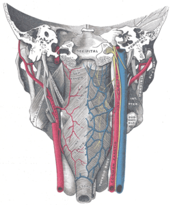
 Section of the neck at about the level of the sixth cervical vertebra. Showing the arrangement of the fascia coli. Carotid sheath is labeled in red. | |
| Details | |
| Identifiers | |
| Latin | vagina carotica fasciae cervicalis |
| TA | A04.2.05.007 |
| FMA | 46561 |
The carotid sheath is an anatomical term for the fibrous connective tissue that surrounds the vascular compartment of the neck. It is part of the deep cervical fascia of the neck, below the superficial cervical fascia meaning the subcutaneous adipose tissue immediately beneath the skin.
The deep cervical fascia of the neck includes four parts:
- The investing layer (encloses the SCM and Trapezius)
- The carotid sheath (encloses the vascular region of the neck)
- The pretracheal fascia (encloses the visceral region of the neck)
- The prevertebral fascia (encloses the vertebral region of the neck)
Structure
The carotid sheath is located at the lateral boundary of the retropharyngeal space at the level of the oropharynx on each side of the neck and deep to the sternocleidomastoid muscle, extending from the base of the skull to the first rib and sternum.
Contents
The four major structures contained in the carotid sheath are:
- the common carotid artery as well as the internal carotid artery
- internal jugular vein
- the vagus nerve (CN X)
- the deep cervical lymph nodes
The carotid artery lies medial to the internal jugular vein, and the vagus nerve is situated posteriorly between the two vessels.
In the upper part, the carotid sheath also contains the glossopharyngeal nerve (IX), the accessory nerve (XI), and the hypoglossal nerve (XII), which pierce the fascia of the carotid sheath.
The ansa cervicalis is embedded in the anterior wall of sheath. It is formed by "descendens hypoglossi" (C1) and "descendens cervicalis" (C2-C3).
Layers
The three major fascial layers in the neck contribute to the carotid sheath: the investing fascia, the pretracheal fascia, and the prevertebral fascia.
Relations
The cervical part of the sympathetic trunk is embedded in prevertebral fascia immediately posterior to the sheath.
See also
Additional images
-

Hypoglossal nerve, cervical plexus, and their branches.
-
Muscles of the pharynx, viewed from behind, together with the associated vessels and nerves.
External links
- MedEd at Loyola grossanatomy/dissector/labs/h_n/pharynx/ph2_1a.html
- lesson8 at The Anatomy Lesson by Wesley Norman (Georgetown University) (latpharyngealitmes)
- MedicalMnemonics.com: 669
- Cross section at tufts.edu